Return to the Histology Tutorial menu.
 Return to the Histology Tutorial menu.
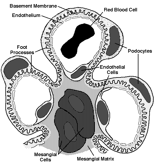
KidneyThe kidney is covered by a connective tissue capsule. The outer part of the kidney is the cortex and the inner part is the medulla. Within the cortex are glomeruli and tubules. There is a grossly and histologically visible corticomedullary junction. Each kidney is supplied with blood by a renal artery, entering at the hilum, which branches into interlobar arteries that branch into the arcuate arteries that run parallel to the corticomedullary junctions. The arcuate arteries give rise to the interlobular arteries of the cortex. Blood from capillaries drains back into the renal vein at the hilum. The functional unit of the kidney is the nephron, which consists of a glomerulus from which a proximal convoluted tubule leads to a thin loop of Henle and then to a distal convoluted tubule. There are about 2 million nephrons per kidney. 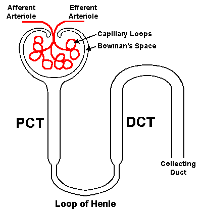
The juxtaglomerular apparatus (JGA) is an important structure that helps to adjust blood volume. There is a region of specialized smooth muscle cells (JG cells) in the afferent arteriole which, along with a set of columnar cells called the macula densa in the adjacent segment of distal convoluted tubule, sense changes in blood pressure and sodium concentration. When blood pressure falls or sodium concentration is reduced, the JG cells of the JGA produces renin. Renin then converts angiotensinogen to angiotensin I, which is then converted to angiotensin II in the lung. Angiotensin II causes arteriolar constriction throughout the body, which raises blood pressure. Angiotensin II also stimulates aldosterone release by the adrenal. The collecting ducts converge into medullary rays that extend into the medulla. These form a renal papilla. Urine drains from the ducts in the papilla into a renal calyx. Each calyx is lined by transitional epithelium. The calyces merge into the renal pelvis. The renal pelvis narrows at the ureteropelvic junction to join the ureter. In the developing fetal kidney, there is a prominent nephrogenic zone from which the cortex is arising. UretersThe ureters run from the ureteropelvic junctions to the ureteral orifices at the bladder trigone. The ureters have an inner longitudinal and outer circular layer of smooth muscle. In addition, the lower third of the ureters has another outer longitudinal layer of smooth muscle. This arrangement helps to produce a continuous peristaltic wave to conduct the urine into the bladder. The ureters have a thin lamina propria beneath the transitional epithelium that lines the lumen. BladderThe ureters enter the bladder at an angle at the trigone, producing a one-way valve effect to prevent reflux of urine upward. The ureteral orifices are located at the bladder trigone. The bladder is an expandable storage container for urine. The bladder has a thick wall composed of branching bundles of smooth muscle that contract to empty the urine. There is a transitional epithelial lining, beneath which is a lamina propria. The bladder epithelium produces a mucoid secretion with natural anti-bacterial properties. This feature, along with normal complete emptying of the bladder, helps to prevent infection. UrethraThe urethra exits the bladder below the trigone. In the male, there is a prostatic portion of the urethra below the bladder that extends through the prostate, and below this, the urethra traverses the urogenital diaphragm before running through the penis (penile urethra) to the urethral meatus at the tip of the glans penis. In a woman, the urethra is much shorter, traversing only the urogenital diaphragm. From the urethral meatus to a variable distance above the urogenital diaphragm, the urethra is lined by stratified squamous epithelium. Above that, there can be a lining of transitional epithelium. |

 |