OBJECTIVE:
- Apply your knowledge of neoplastic, inflammatory, and developmental diseases to interpret clinical history, laboratory tests, and pathologic findings for diagnosis of lesions of the head and neck region.
CASE 1
(Click here to go to the answers)
Clinical History:
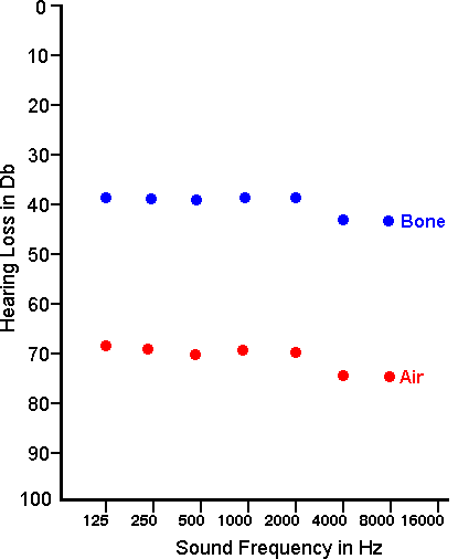
- A 9-year-old boy has had difficulty hearing on the right. As a child, he had multiple episodes of otitis media. This progressed to chronic otitis media on the right, with persistent drainage, and the tympanic membrane ruptured when he was a teenager. Since that time hearing in his right ear has been diminished. His audiogram on the right is shown here:
Questions:
- What are common infectious agents that produce acute otitis media?
- What type of hearing loss is he most likely to have?
- How is serous otitis media caused in children?
Further History:
- A computed tomographic (CT) scan shows a 2 cm mass involving the right middle ear and mastoid (image 1.1). The mass is surgically excised. The microscopic appearance of the mass is shown in images 1.2 and 1.3. What are the microscopic findings?
-
-
-
Questions:
- What is the name for this mass lesion?
- How does this lesion occur?
- Name a cause for progressive hearing loss in the elderly
|
CASE 2
(Click here to go to the answers)
Clinical History:
- A 39-year-old woman has noted persistent headaches and dizziness for the past 4 months. She also notes diminished hearing on the left. Audiometry reveals the findings shown here:

-
- MR imaging is performed, followed by surgery.
Radiology and Microscopic Pathology:
- The appearance of the lesion with MRI is shown here (image 2.1). Describe the histologic findings (images 2.2 to 2.3).
-
-
-
Questions:
- Where is this mass located?
- What is the diagnosis?
- What is the prognosis?
- Name a condition in which this lesion can be seen in association with multiple neoplasms.
|
CASE 3
(Click here to go to the answers)
Clinical History:
- A healthy 18-year-old has a painful lesion on his lower lip that has been present for several days following a backpacking trip to climb Mt. St. Elias. He employs aromatherapy (a kit for less than $20 US purchased at a local shopping mall) and the lesion resolves in two weeks.
Gross and Microscopic Pathology:
- The appearance of the lesion is shown here. How would you describe it? (images 3.1 to 3.3)
-
-
-
Questions:
- What is the diagnosis?
- What is the cause of this lesion.
- What is in the differential diagnosis of this condition?
- What is the prognosis?
- What therapies are available?
|
CASE 4
(Click here to go to the answers)
Clinical History:
- A 72-year-old man is bothered by headaches, increasing in frequency and severity over the past month. He has some facial pain on the left. There is photophobia on the left along with decreased vision.
Gross amd Microscopic Pathology:
- Here is the gross appearance of this forehead on the left. What is the abnormality? (image 4.1). The microscopic appearance of the lesion at low and high power is shown here. What are the findings? (images 4.2 and 4.3).
-
-
-
Questions:
- What laboratory test would you order?
- What is the diagnosis?
- How is this condition treated?
- What systemic condition may accompany this lesion?
|
CASE 5
(Click here to go to the answers)
Clinical History:
- A 60-year-old man has increasing hoarseness over the last 6 months. He has smoked 2 packs of cigarettes per day for the past 45 years, and has had a chronic cough for may years. However, yesterday he noted some blood streaking of his sputum. Endoscopy reveals a mass invoving the vocal cords, right more than left. He has some enlarged non-tender cervical lymph nodes.
Radiologic, Gross, and Microscopic Pathology:
- What is the location (image 5.1)? What does the gross appearance suggest (image 5.2)? What is seen microscopically (image 5.3)?
-
-
-
Questions:
- What is the diagnosis?
- What is the risk factor for this condition?
- What is the prognosis?
- What is palliative care?
|
CASE 6
(Click here to go to the answers)
Clinical History:
- A 12-year-old child has complained of a "bump" on his neck for the past month. Physical examination reveals a nontender mass about 2 cm in size in the midline of the neck beneath the skin above the thyroid cartilage. A head and neck CT scan is performed. The mass is excised and examined in surgical pathology.
Radiographic, Gross, and Microscopic Pathology:
- The head CT scan is shown in image 6.1). The gross appearance at surgery is seen in image 6.2. The microscopic appearance is seen in image 6.3.
-
-
-
Questions:
- What is the diagnosis?
- Explain how this lesion occurs.
- What is the differential diagnosis of this lesion?
|
CASE 7
(Click here to go to the answers)
Clinical History:
- A 43-year-old woman has noticed a painless swelling on the left side of her face, increasing for over 2 years. When examined by her physician there is a 4 cm firm, mobile mass anterior to the lower left ear. The mass is excised.
Findings:
- The appearance of the mass with MR imaging is shown (image 7.1). The low and high microscopic appearances of the mass are shown here (images 7.2 and 7.3).
-
-
-
Questions:
- What is the diagnosis?
- What is the prognosis?
- What is the differential diagnosis for this mass?
|
CASE 8
(Click here to go to the answers)
Clinical History:
- A 39-year-old man has been bothered by nasal congestion with watery eyes and sneezing every spring and summer for many years. He now has increasing difficulty breathing through his nose. Examination of his nose reveals several 1 to 2 cm glistening masses occluding each nostril. These masses are excised.
Microscopic Pathology:
- The appearance of the nasal cavity and paranasal sinuses is seen with CT scan (image 8.1). The low and high power microscopic appearance of the masses are shown here. (images 8.2 and 8.3)?
-
-
-
Questions:
- What is the diagnosis?
- What is the pathogenesis?
|
|
 Return to the
Laboratory Menu.
Return to the
Laboratory Menu.