 Return to the Histology Tutorial menu.
 Return to the Histology Tutorial menu.
|
The two major divisions of the nervous system are the central nervous system (CNS), consisting of the brain and spinal cord, and the peripheral nervous system (PNS) consisting of the peripheral nerves, nerve receptor endings, and ganglia distributed throughout the body. Embryologically, the central nervous system is derived from the neural plate which closes to form the neural tube. From the anterior neural tube additional folding and development produces the complex regions of the brain. The neural crest cells migrate to give rise to most of the peripheral nervous system. Cells of the Nervous System - NeuronsThe major function of the nervous system is provided by neurons. Neurons come in a variety of shapes and sizes. In general, they have the following components: 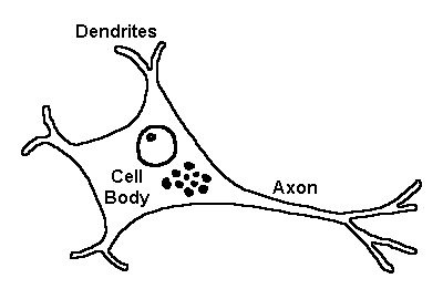
Neurons can be subdivided into:
Cells of the Nervous System - Glial CellsThere are a variety of cells which support neuronal function. These include:
Structural Components and RegionsThese cells form the substance of the brain, spinal cord, and peripheral nervous system. The brain is composed of the basic units of the cerebral hemispheres, brainstem, and cerebellum. The cerebral hemispheres can be further categorized by structure and function. Grossly, structures such as the cerebral cortex have many neurons and appear grey, the so-called "grey matter." Regions of the brain that are composed primarily of fiber tracts making connections appear lighter, the so-called "white matter". Histologically, the cortex of the cerebral hemispheres can be categorized as "neocortex" with six layers, considered more developed, and the "paleocortex" with three layers, seen in central structures controlling more primitive reflexive, emotional functions of the limbic system, such as the hippocampus. The hippocampus is also involved in processing short term memories. The cerebellum is involved with controlling and fine-tuning movement. There are three layers comprising the cerebellar cortex. The spinal cord is arranged with outer white matter and inner grey matter with neurons such as the anterior horn cells. From the spinal cord emanate the spinal nerve roots. MeningesThe central nervous system is covered by specialized connective tissues that are divided into three layers:
Ependymal Lining and Choroid PlexusesThe cerebral ventricles and spinal canal containing the cerebrospinal fluid are lined by an ependyma, which consists of a single layer of cuboidal to columnar cells that have cilia or microvilli. In the lateral ventricles and the fourth ventricle, the ependyma is specialized to form the choroid plexuses. These plexuses consist of a highly vascularized, arborizing stroma covered by cuboidal cells with fine microvilli. The choroid plexus produces approximately 0.5 to 1.5 liters of cerebrospinal fluid per day. This CSF is an ultrafiltrate of plasma that provides a "shock absorber" function for the brain. The CSF circulates through the ventricles and spinal canal. The CSF is reabsorbed at the arachnoid granulations at the vertex of the brain. If the flow of CSF is blocked, a condition known as hydrocephalus can occur. Blood Brain Barrier (BBB)The BBB is a diffusion barrier that prevents the influx of most compounds from blood to brain. The three cellular elements of the BBB are endothelial cells, astrocytic foot processes, and pericytes (PCs). Pericytes (PCs) encircle endothelial cells and provide structural support and aid in controlling blood flow. The tight junctions (TJs) between cerebral endothelial cells form the selective diffusion barrier. Astrocytic foot processes closely adhere to small vessels and aid in the induction and maintenance of the TJ barrier. However, the astrocytes do not have a direct barrier function. Loss of the astrocytes leads to downregulation of TJ proteins such as occludin and claudin, with resultant diffusion of some, but not all, proteins through the endothelial cells. The BBB is present in all brain regions, except for certain areas that regulate autonomic nervous system functions and endocrine functions of the body such as the pituitary. Fenestrations in blood vessels in these select areas permit diffusion of molecules across the vessel wall. Peripheral Nervous SystemThe PNS is comprised of several structures. There are ganglia that are formed of a group of cell bodies. The Schwann cells are specialized to form satellite cells that invest the cell bodies. The dorsal root ganglia next to the spinal cord are involved with sensory function. The autonomic nervous system has autonomic ganglia that can be further divided into sympathetic and parasympathetic ganglia. The axons of neurons are typically grouped together to form nerve trunks. There are connective tissue layers that surround the peripheral nerves. An endoneurium surrounds an individual axon. Bundles of axons called fascicles are surrounded by a perineurium. The largest nerve trunks composed of multiple fascicles are surrounded by an epineurium. The peripheral nervous system has ganglia that consist of localized collections of nerve cell bodies surrounded by a connective tissue capsule. A ganglion provides a location for synapses. There are dorsal root ganglia adjacent to the spinal cord. There are ganglia associated with the autonomic nervous system known as parasympathetic and sympathetic ganglia. The actual sensory input into the periphral nervous system comes from a variety of specialized nerve endings and receptors. The sense of touch is one example. One of the largest, most impressive receptors is the pressoreceptor, or Pacinian corpuscle. |

 |