Combinations of Hemoglobin S, C, E, and beta thalassemia
These variants arose in different geographic regions, and represent different alterations in beta globin chains. Combinations of these different defects are possible. Findings with hemoglobin electrophoresis are helpful in distinguishing them.
Hemoglobin SC Disease
In this disorder, which is most often found in West Africa, there is one beta globin gene coding for hemoglobin S and the other coding for hemoglobin C. The result is a disease that is milder than sickle cell anemia, but worse than C trait. Affected persons may have a mild to moderate anemia with some sickling and hemolysis. Onset in childhood is not usually accompanied by serious complications, but teenagers and adults may have recurrent infections, joint and muscle pain with crises, and icterus. Aseptic necrosis of the femoral head may occur. Vaso-occlusive "crises" are rare.
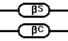
A peripheral blood smear will demonstrate many target cells and peculiar "SC poikilocytes". Elongated hexagonal "SC crystals" may be found in some RBC's. Occasional sickle cells can be present. The sickle test will be positive. Hemoglobin electrophoresis will reveal that about half hemoglobin S and half hemoglobin C.
The following images illustrate pathologic findings with hemoglobin SC disease:
Hemoglobin S/beta-0 thalassemia
Persons with this combination have one hemoglobin S gene and one beta-0 thalassemia gene. This disorder is nearly as bad as sickle cell anemia. There is risk for vaso-occlusive crises. Unlike sickle cell anemia, there is usually splenomegaly.
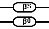
A peripheral blood smear will demonstrate microcytosis, some hypochromic RBC's, and some target cells. Hemoglobin electrophoresis will reveal 90 to 95% hemoglobin S, 5% hemoglobin A2, and 5 to 10% hemoglobin F.
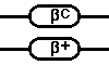
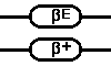
Hemoglobin S/beta+ thalassemia
This combination is better than sickle cell anemia. Hemoglobin electrophoresis demonstrates 5 to 30% hemoglobin A, 60 to 90% hemoglobin S, 5% hemoglobin A2, and 5 to 10% hemoglobin F. In contrast to sickle cell trait (Hgb AS), the amount of hemoglobin S is greater than the amount of hemoglobin A.

Hemoglobin C/beta thalassemia
In this disorder, there can be a mild to moderate anemia, depending upon whether the beta-0 or beta+ variant is present. Hemoglobin electrophoresis will demonstrate 60 to 95% hemoglobin C, 1 to 5% hemoglobin F, and 0 to 20% hemoglobin A (none with beta-0 variant)
 OR
OR 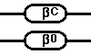
Hemoglobin E/beta thalassemia
The findings in this condition resemble beta-0 thalassemia major. The results of hemoglobin electrophoresis are quite variable and will demonstrate 15 to 95% hemoglobin E, 5 to 85% hemoglobin F, and virtually no hemoglobin A.
 OR
OR 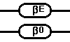
 Next
Next Back
Back