Hemoglobin C and Hemoglobin E Disease
These two variants arose in different geographic regions, and represent two different alterations in beta globin chains, owing to single amino acid substitutions. Hemoglobin electrophoresis is helpful in distinguishing these variants.
Hemoglobin C Disease
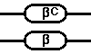
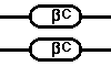
In this disorder, which appears to have arisen in West Africa, there is a single amino acid substitution in the beta globin chain. Just like sickle cell disease, the substitution occurs at position six, but lysine substitutes for glutamic acid. The result is hemoglobin C that does not lead to sickling, but to abnormal RBC's that appear as "target cells". When hemoglobin C is present, the amount of hemoglobin A2 is difficult to assess, as these two hemoglobins are hard to distinguish via electrophoretic methods.
Persons heterozygous for the C gene will have no significant problem, and a peripheral blood smear will reveal the presence of occasional target cells. Hemoglobin electroporesis will demonstrate about 40 to 50% hemoglobin C and 50 to 60% hemoglobin A.

Persons homozygous for the C gene will have a mild hemolytic anemia with indirect hyperbilirubinemia and splenomegaly. A peripheral blood smear will reveal many target cells. The MCV will be low (with resultant microcytosis) but the MCHC will be high because of the presence of spherocytes. Elongated hexagonal crystals may appear in some of the cells, particularly in splenectomized patients. Hemoglobin electrophoresis will demonstrate 90 to 95% hemoglobin C and 1 to 7% hemoglobin F.
Hemoglobin E Disease
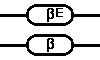
The abnormal beta globin gene for this disorder arose in Southeast Asia. There is an abnormal amino acid substitution on the globin chain at position 26, with lysine substituted for glutamic acid. This abnormal hemoglobin does not lead to sickling, and RBC's containing this abnormal hemoglobin E have only the tendency to exhibit microcytosis.
Persons heterozygous for the E gene will have no anemia, and the RBC's will have decreased MCV, with microcytosis as a consequence. Hemoglobin electrophoresis will demonstrate 30 to 40% hemoglobin E and 60 to 70% hemoglobin A.
Persons homozygous for the E gene will have mild anemia and RBC's with lower MCV (microcytosis). Hemoglobin electrophoresis will demonstrate 90 to 95% hemoglobin E, 3 to 5% hemoglobin A2, and 1 to 5% hemoglobin F.
 Next
Next Back
Back