Cardiac Physiology
Return to the Main Menu
Case 1:
(Click here to go to the answers)
A 68-year-old woman has noted increasing difficulty with breathing for the past 4 years. She has become increasingly fatigued. She has had episodes during the past 6 months of waking up at night with marked shortness of breath, coughing up frothy sputum.
A chest radiograph shows cardiomegaly, blunting of the costophrenic recesses, prominent pulmonary arteries, and peripheral lower lobe opacifications.
On physical examination vital signs include T 37.0 C, P 82/min, RR 20/min and labored (but without use of accessory muscles), and BP 155/90 mm Hg. She has poor peripheral capillary filling and cold hands. There is pitting edema to the knees. There is jugular venous distension while sitting to the angle of the jaw. Auscultation of the chest shows a holosystolic murmur and diminished S1 and S2. Diffuse bilateral coarse inspiratory crackles are heard over both lung bases. Her liver edge is palpable, but the spleen is not palpable.
Laboratory studies show Na 139 mmol/L, K 3.9 mmol/L, Cl 101 mmol/L, HCO3 28 mmol/L, glucose 124 mg/dL, creatinine 1.4 mg/dL, and urea nitrogen 25 mg/dL.
An EKG is performed. Review the features of a normal EKG.
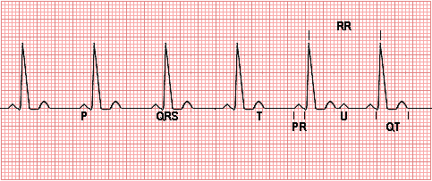
Each small square represents 0.04 second horizontally and 0.01 millivolt vertically.
The P wave represents depolarization of the atria.
The QRS complex represents bilateral ventricular depolarization. The QRS duration is normally 0.06 - 0.10 seconds and represents the duration of ventricular muscle depolarization.
The PR interval is the time interval from the onset of atrial depolarization (P wave) to the onset of ventricular depolarization (QRS complex). The normal PR interval is 0.12 - 0.20 seconds.
The ST-T wave represents ventricular repolarization.
The QT interval (QTc) represents the duration of ventricular depolarization and repolarization and is normally < 0.40 seconds at a heart rate of 70/min or above.
The U wave, if present, represents "after depolarizations" in the ventricles.
The RR interval is the duration of the ventricular cardiac cycle, an indicator of the ventricular rate.
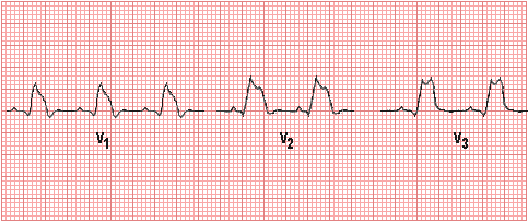
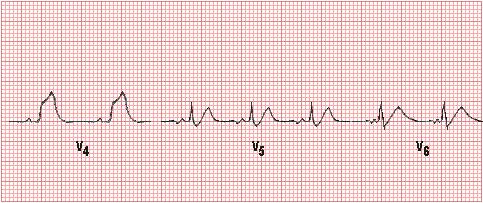
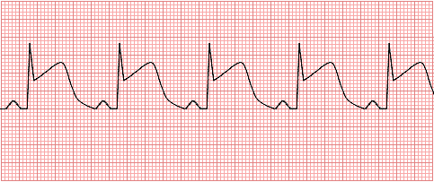
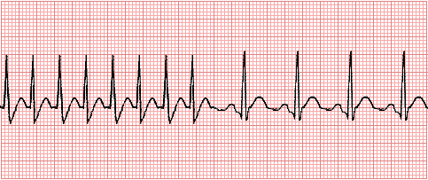
An EKG is performed in this patient which shows a regular rate and rhythm but increased amplitude in lead II.
Cardiac catheterization:
- Chamber volume at end of diastole = 0.138 L
- Chamber volume at end of systole = 0.083 L
- Heart rate = 91/min
- Left ventricular end-diastolic pressure 12 mmHg
- Pulmonary arterial pressure 50/20 mm Hg, mean 30 mmHg
- Pulmonary artery wedge pressure = 20 mm Hg
- Right atrial pressure = 12 mm Hg
- Coronary arteries show <20% stenosis
Questions:
1.1 What is her stroke volume?
1.2 What is her cardiac output?
1.3 What is her cardiac index (her body surface area is 1.6 m2)?
1.4 What is her ejection fraction?
The ejection fraction (EF) is the amount of blood ejected from the left ventricle (LV) with each heart beat and is normally > 55%. An EF can be calculated with an echocardiogram, a left ventriculogram done during a cardiac catheterization, or a nuclear study called a MUGA. The method of calculating the EF involves tracing the dimensions of the LV at the end of its contraction period (systole) and at the end of its relaxation period (diastole). These dimensions are then used to calculate the EF with a standard mathematical formula. Many cardiologists visually estimate the EF. Generally, the calculated EF is more accurate. The lower the EF, the worse the congestive heart failure.
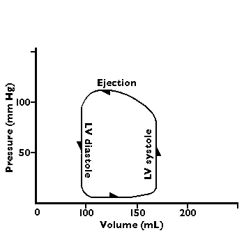
A flow-volume loop diagram of normal cardiac output has the following appearance:
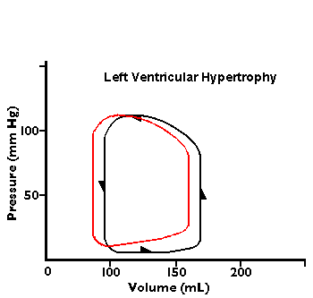
In the diagrams below, abnormal flow-volume loops are shown:

1.5 Compare these cardiac catheterization findings to normal.
1.6 What determines the voltage that is recorded on the limb leads? Mention several factors.
1.7 What sorts of things lengthen the PR interval? Can you suggest how they might work?
1.8 Does she have right or left heart disease or both?
1.9 What cardiac problem is causing these findings?
1.10 What therapies are available?
1.11 If the patient had a blood pressure of 165/100 mm Hg, what pharmacologic therapy would you consider?
Case 2:
(Click here to go to the answers)
A 39-year-old man comes to the emergency department with chest pain. The marked pain had an onset 4 hours ago and has not abated. He has not experienced a similar pain in the past. This pain is not associated with breathing or swallowing movements. On physical examination, vital signs show T 37.1 C, P 100/min, RR 19/min, and BP 100/65 mm Hg. His skin is cool, with normal turgor. His lungs are clear to auscultation. His heart rate is slightly irregular. No murmurs are heard. Peripheral pulses are 1+ in lower and upper extremities. His abdomen is non-tender and bowel sounds are present. The stool guaiac is negative for occult blood. No neurologic problems are present.
Initial laboratory findings: hematocrit 40%, glucose 80 mg/dL, dipstick urinalysis normal.
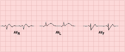
Electrocardiogram:

Questions:
2.1 What is your initial impression?
Review the probable progression of EKG findings seen with acute MI:
1: Normal ECG prior to MI
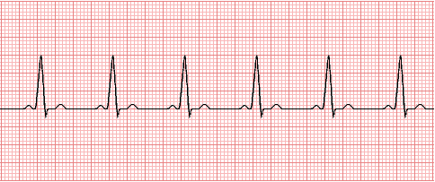
2. Early ischemia with increased T wave amplitude and width; minimal ST segment elevation
3. More extensive ischemia with marked ST segment elevation and T wave changes
4. Early infarction with pathologic Q wave, less ST elevation, and terminal T wave inversion
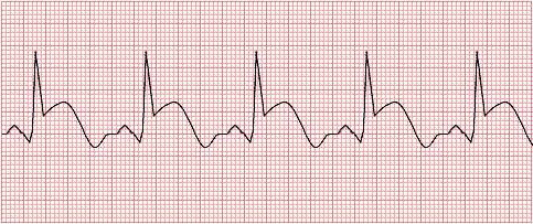
5. Healing infarction (1 to 2 weeks) with pathologic Q wave and T wave inversion
6. Healing infarction (1 month) with residual small pathologic Q wave and upright T wave
2.2 What additional laboratory test finding is most likely to be present?
2.3 What laboratory test(s) would you get later, when is is more stable, for risk assessment?
2.4 What pharmacologic therapy would you consider at this point?
2.5 If his total serum cholesterol is 262 mg/dL, with serum triglyceride 177 mg/dL, and glucose 111 mg/dL, what therapy would you consider, and what would be the mechanism of action and potentially the most serious adverse effects?
2.6 How is his cardiac output and stroke volume likely to be altered?
2.7 What is the effect on coronary blood flow of a 50% narrowing?
Case 3:
(Click here to go to the answers)
A 72-year-old man has the sudden onset of severe abdominal pain. He is taken to the emergency department. On admission his vital signs show T 37.1 C, P 95/min, RR 18/min, and BP 70/40 mm Hg. Peripheral pulses are weak. His skin is cool and clammy. Auscultation of the chest shows lung fields are clear. His heart rate is regular with no murmurs. His abdomen is diffusely and markedly tender with no bowel sounds present. His stool is negative for occult blood.
Initial laboratory studies show hematocrit 17%, sodium 140 mmol/L, potassium 4.0 mmol/L, chloride 105 mmol/L, CO2 26 mmol/L, glucose 71 mg/dL, creatinine 1.1 mg/dL, and amylase 43 U/L.
An abdominal CT scan shows a calcified abdominal aorta with diameter of 6 cm. There is an accumulation of fluid in the abdomen.
Questions:
3.1 What is the most likely diagnosis?
3.2 What therapeutic measures are indicated?
Case 4:
(Click here to go to the answers)
An 80-year-old woman has been bothered for the past 3 months by what she describes as a "fluttering" of her heart that happens about once per week, lasting for about 10 minutes. The episodes seem to go away if she drinks a glass of blackberry brandy. She has had no medical problems in the past. On physical examination there are no remarkable findings.
Laboratory findings show: hematocrit 43%, glucose 87 mg/dL, total cholesterol 182 mg/dL, creatinine 0.7 mg/dL, and troponin I <1 ng/mL.
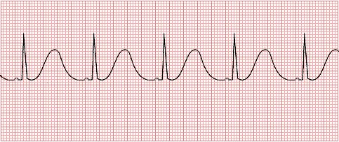
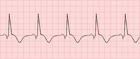
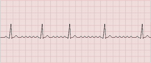
An EKG shows the following:
Questions:
4.1 What is the most likely diagnosis?
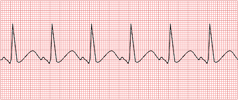
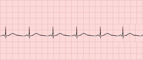
Compare with the following EKG:
4.2 What is the cardiac abnormality?
4.3 What pharmacologic therapy is indicated?
4.4 What is the effect of the blackberry brandy?
4.5 What is the internal pacemaker of the heart and what is its natural rate? What is the effect of sympathetic or parasympathetic stimulation?
Case 5:
(Click here to go to the answers)
A 10-year-old boy was riding his bike and fell, hitting his head on the ground. His mother found him unconscious, with a bloody forehead. She thought he was dead, but the boy regained consciousness before she could phone 911. She brings the boy to the emergency department. He has a small laceration to the forehead, which requires 5 sutures. A week later, she finds him collapsed on the ground, and he regains consciousness within 5 minutes. A trip to the emergency department yields no findings on physical examination. This scenario is repeated 4 more times in the next 6 months. Each time a different physician is on duty in the emergency department, and they have to repeat the historical findings each time. Her husband, having returned from a tour of military duty, accompanies her with the child on the 7th ED visit. He remarks that his Aunt Emma had similar episodes before she died suddenly at age 39.
Questions:
5.1 What conditions are in your differential diagnosis?
5.2 What test(s) or procedure(s) would be helpful?
What is shown here?
5.3 What complication is shown here?

5.4 What causes this finding?
5.5 What can be done?
Case 6:
(Click here to go to the answers)
A 75-year-old man visits an optometrist due to diminishing vision. The optometrist performs funduscopy and recognizes features of hypertensive retinopathy. He tells the man to visit a physician. On physical examination, vital signs include temperature 37 C, pulse 75/minute, respiratory rate 16/minute, and blood pressure 170/110 mm Hg. The remainder of the physical examination is unremarkable except for reduced range of motion at the knees and hips. His BMI is 28.
A chest radiograph shows a prominent left heart border along with calcification of the aortic knob. Laboratory studies show Hgb 13.5 g/dL, Hct 40.4%, MCV 80 fL, platelet count 186,000/microliter, WBC count 6380/microliter, serum sodium 143 mmol/L, potassium 4.2 mmol/L, chloride 105 mmol/L, CO2 24 mmol/L, glucose 128 mg/dL, creatinine 1.2 mg/dL, triglyceride 177 mg/dL, and total cholesterol 192 mg/dL.
Questions:
6.1 What is the diagnosis and what are risk factors?
6.2 Explain the physiology of his condition.
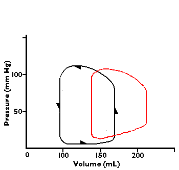
The effect upon the heart from increased afterload is shown below:

6.3 In what condition would systolic pressure be reduced, but diastolic pressure be maintained?
6.4 What pharmacologic therapy is indicated for this man? Find specific agents and dosing regimens that the patient could reasonably follow, given that you have to avoid drug interactions or adverse events with his current medications, which include:
- Insulin
- Ibuprofen
- Allopurinol
- Cimetidine
- Warfarin
- Ginkgo biloba
Return to the Main Menu
|