|
A 39-year-old man comes to the emergency department with chest pain. The marked pain had an onset 4 hours ago and has not abated. He has not experienced a similar pain in the past. This pain is not associated with breathing or swallowing movements. On physical examination, vital signs show T 37.1 C, P 100/min, RR 19/min, and BP 100/65 mm Hg. His skin is cool, with normal turgor. His lungs are clear to auscultation. His heart rate is slightly irregular. No murmurs are heard. Peripheral pulses are 1+ in lower and upper extremities. His abdomen is non-tender and bowel sounds are present. The stool guaiac is negative for occult blood. No neurologic problems are present.
Initial laboratory findings: hematocrit 40%, glucose 80 mg/dL, dipstick urinalysis normal.
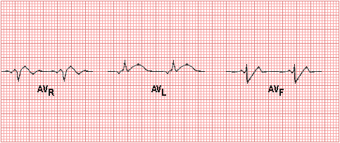
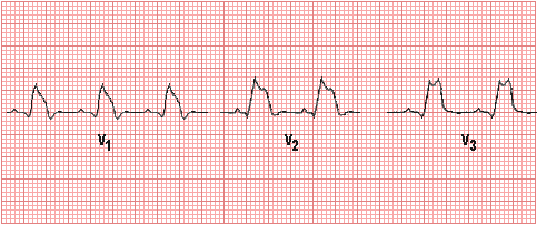
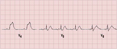
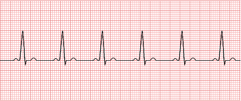
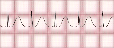
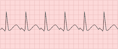
Electrocardiogram:

Questions:
2.1 What is your initial impression?
The rate and rhythm of the EKG are normal. The PR interval is normal. There are pathologic Q waves in leads V1-V4, and in the precordial leads there is loss of R wave progression.
He has an acute coronary syndrome.
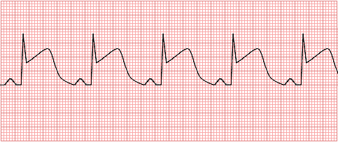
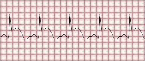
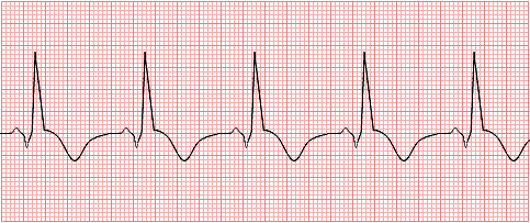
Review the probable progression of EKG findings seen with acute MI:
1: Normal ECG prior to MI
2. Early ischemia with increased T wave amplitude and width; minimal ST segment elevation
3. More extensive ischemia with marked ST segment elevation and T wave changes
4. Early infarction with pathologic Q wave, less ST elevation, and terminal T wave inversion
5. Healing infarction (1 to 2 weeks) with pathologic Q wave and T wave inversion
6. Healing infarction (1 month) with residual small pathologic Q wave and upright T wave
2.2 What additional laboratory test finding is most likely to be present?
The troponin I or the CK-MB may be elevated. An elevation in either of these markers of cardiac injury may appear starting 3 hours from the onset of myocardial ischemia.
2.3 What laboratory test(s) would you get later, when is is more stable, for risk assessment?
A coronary risk panel with markers for hyperlipidemia can assess his underlying risk for atherosclerotic cardiovascular disease. A fasting serum glucose can screen for diabetes mellitus.
2.4 What pharmacologic therapy would you consider at this point?
Thrombolytic therapy (with tPA) may be considered. Post-MI, beta blockers are usually beneficial, but inotropic agents are generally not helpful.
2.5 If his total serum cholesterol is 262 mg/dL, with serum triglyceride 177 mg/dL, and glucose 111 mg/dL, what therapy would you consider, and what would be the mechanism of action and potentially the most serious adverse effects?
Atorvastatin (Lipitor) and rosuvastatin (Crestor) are the most effective. These drugs are competitive inhibitors of HMG-CoA reductase that converts 3-hydroxy-3-methylglutaryl coenzyme A to mevalonate, a precursor of cholesterol, thus reducing endogenous cholesterol synthesis in the liver. These "statins" also increase the number of hepatic LDL receptors to enhance uptake and catabolism of LDL cholesterol. Their most serious adverse effect is rhabdomyolysis with acute renal failure secondary to myoglobinuria, which has been reported with drugs in this class. Liver dysfunction has also been reported, and patients with a significant (>3X) rise in transaminases should stop the drug.
If statins are not tolerated or do not produce sufficient lipid lowering effect, then ezetimibe (Zetia) may be considered. Ezetimibe reduces blood cholesterol by inhibiting the absorption of cholesterol by the small intestine, but without significant effect upon absorption of nutrients such as fat soluble vitamins.
2.6 How is his cardiac output and stroke volume likely to be altered?
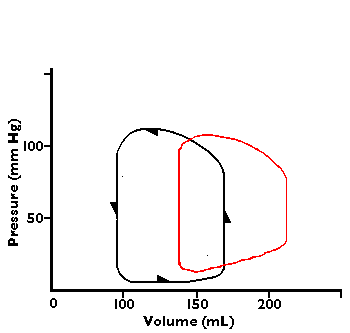
There is a decreased stroke volume and decreased cardiac ouput from loss of myocardial contractility involving the left ventricle.
2.7 What is the effect on coronary blood flow of a 50% narrowing?
If the radius of the artery is reduced to half, the resistance increases 16 fold. Since flow is inversely proportional to resistance, then the flow is reduced to 1/16. Thus, narrowing has a marked effect on blood flow.
| 
