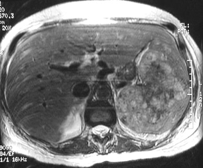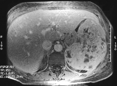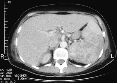
| There is a heterogenous splenic mass seen in the abdominal CT scan above. This large mass expands the spleen. Histologically, it was an angiosarcoma. In the T1 weighted MRI scan below can be seen areas of increased signal intensity characteristic for hemorrhage in a vascular neoplasm. A T2 weighted MRI scan is shown at the bottom in which the hemorrhagic-vascular areas appear darker. |
|
|