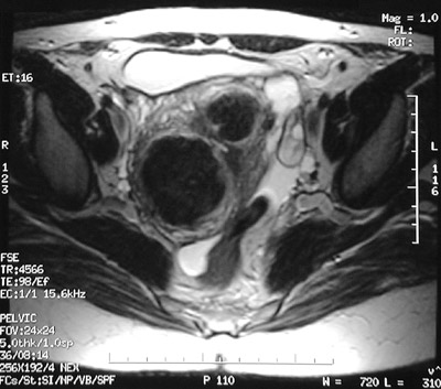
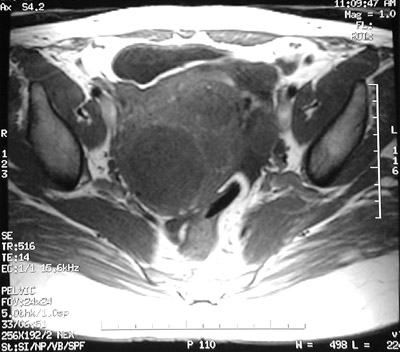
These axial MRI scans of the pelvis show a large nodular uterus containing leiomyomata. The T2 weighted FSE image above and the T1 weighted SE image below both reveal a
larger
as well as a
smaller
mass involving the uterus.