


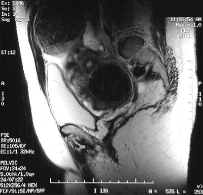
| This T2 weighted FSE MRI scan of the pelvis in sagittal view reveals a large nodular uterus containing leiomyomata. One large leiomyoma is seen protruding subserosally from the upper fundus, while another leiomyoma is seen intramurally in the lower portion of the uterus compressing the bladder anteriorly. |
|
|