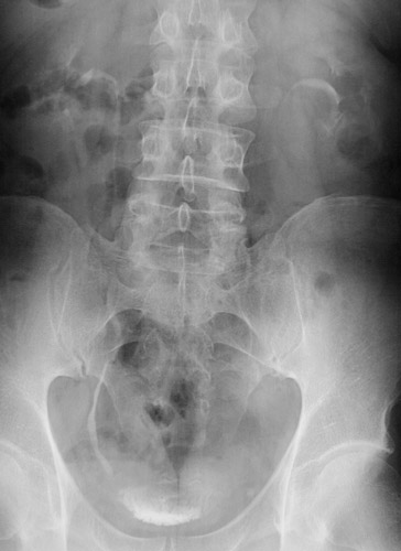
This intravenous pyelogram demonstrates a
ureteral calculus
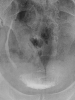
at the vesicoureteral junction on the left where there is a filling defect. A closer view is seen below.