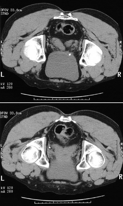
The two CT scan views above in a patient, who is being scanned while prone, contrast a
ureteral calculus
at the vesicoureteral junction with a
phlebolith
seen in soft tissue near the pelvic side wall.