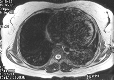
Seen in this axial T2 weighted FSE axial MRI scan of the chest is a
markedly enlarged heart
in a patient with hereditary hemochromatosis. Note the decreased signal intensity of the heart from the increased iron deposition.