


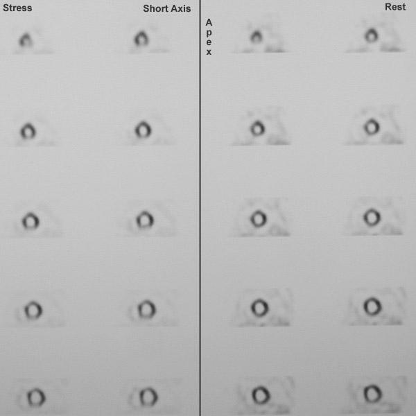
| This is a SPECT (single-photon emission computed tomography) scan that gives the appearance of the heart, principally the left ventricle, in three planes. The "short axis" plane is seen here, beginning at the apex (top of image) and proceeding toward the base. This radioisotopic imaging technique uses thallium. In this study, the patient is undergoing a stress test in which the images at the right were produced within minutes of injection while the subject was exercising. The images at the left were taken later after resting. The images reveal an area of decreased uptake inferiorly that represents ischemic myocardium under stress. This lighter area becomes darker (perfused) while the subject is at rest. |
|
|