


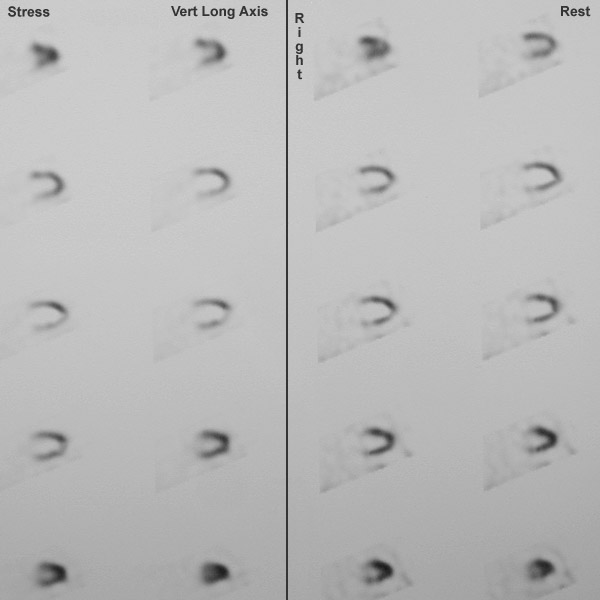
| This is a SPECT nuclear medicine scan that is shown in the "vertical long axis" plane, beginning toward the right (top of image); the superior aspect is the anterior portion of the heart. In this study, the patient is undergoing a stress test in which the images at the right were produced within minutes of injection while the subject was exercising. The images at the left were taken later after resting. The images reveal an area of decreased uptake inferiorly that represents ischemic myocardium under stress. This lighter area becomes darker (perfused) while the subject is at rest. |
|
|