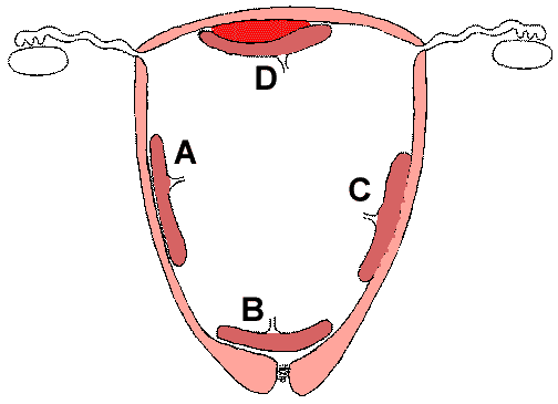
- Placenta previa (B):
- The placenta implants in the lower uterine segment and is in a position to be traumatized (with severe hemorrhage) during the normal vaginal birth process.
- Placenta accreta (C):
- A normal decidual plate is lacking, and the villi implant into the myometrium. Thus the placenta cannot separate normally at delivery and a hysterectomy must be done. This condition is fortunately uncommon.
- Abruptio placenta (D):
- There is abnormal premature separation of the placenta with formation of a blood clot. This compromises the blood supply to the fetus.
- Accessory lobe:
- The placenta may have an accessory lobe that could be retained following delivery.
- Tubal (ectopic) pregnancy:
- The fertilized ovum implants at an abnormal site, most often the fallopian tube. The growth of the embryo leads to pain, hemorrhage, and possible rupture (a medical emergency).
|
PART II - PERINATAL PATHOLOGY
CASE 1
(Click here to go to the answers)
History:
- A 25-year-old primigravida gave birth to a 35 week gestational age male infant. The baby had Apgar scores of 4 at 1 minute and 6 at 5 minutes. The baby appeared hydropic, as did the placenta. The baby appeared to be in congestive heart failure. Urine output was poor. Despite resuscitative measures, the baby died 2 days after delivery.
-
-
Questions:
Name some causes for hydrops fetalis. What was the prognostic value of the Apgar scores? What is the etiology in this case?
|
CASE 2
(Click here to go to the answers)
History:
- A 23-year-old primigravida had received routine prenatal care throughout her pregnancy and had problems only with mild hyperemesis gravidarum. The size of the fetus by examination was consistent with 38 weeks gestational age on her last prenatal visit. However, a week later she called her physician because she had not felt any fetal movement for a whole day. An ultrasound performed at the physician's office revealed no cardiac activity. A stillborn baby was delivered soon thereafter. The placenta weighed 550 gm and had an 82 cm long umbilical cord with three vessels that inserted paracentrally.
-
-
-
Questions:
What is the significance of umbilical cord length? What determines the length of the cord? What is the significance of the nuchal cord?
|
CASE 3
(Click here to go to the answers)
History:
- A 28-year-old gravida 2 para 0 ab 1 woman has a routine ultrasound at 26 weeks gestation. Very little amniotic fluid is noted, and it is difficult to visualize the fetus. On repeat ultrasound a week later, the same findings are noted, but the kidneys are now visualized and appear to be enlarged and cystic bilaterally. The baby is born prematurely at 30 weeks gestation by spontaneous vaginal delivery and weighs 850 gm. Apgar scores are 1 at 1 minute, 1 at 5 minutes, and 1 at 10 minutes (heart rate only). The baby dies at 1 hour of age.
-
-
-
-
-
-
-
Questions:
Is this early or late onset intrauterine growth retardation? How do you explain the oligohydramnios? Why were the Apgar scores low? How do you explain the pathophysiology of the gross and microscopic
findings? Explain the genetic basis for the conditions in images 3.5 and 3.6 for
the purpose of counselling the parents. What is the prognosis and how do you help the parents and family to deal with it?
|
CASE 4
(Click here to go to the answers)
History:
- A 21-year-old primigravida has noted leakage of amniotic fluid for a couple of days and presents to the emergency room with a fever of 38.8 C. On physical examination, there is a foul-smelling vaginal discharge. A culture is taken. The cervix is dilated to 5 cm. An ultrasound reveals a fetus of 28 week gestational age with evidence for cardiac activity. She is in premature labor. Several hours later, a 900 gm male fetus is delivered. The Apgar scores are 5 at 1 minute and 6 at 5 minutes. The baby receives artificial surfactant via endotracheal tube following intubation. The baby is ventilated with PEEP. The following day, the baby has a generalized seizure and cranial ultrasound reveals a massive intraventricular hemorrhage. The baby expires later that day.
-
-
-
Questions:
Is this baby small, appropriate, or large for gestational age? What is the pathophysiology of premature rupture of membranes?
What did the culture probably grow? What is the cause for the pulmonary disease? What is the cause for the intraventricular hemorrhage? Name the major causes for congenital infections. What social and ethical issues are raised with long-term ventilatory support?
|
CASE 5
(Click here to go to the answers)
History:
- These male infants are the product of a 37 week gestation in a 35-year-old mother whose only previous pregnancy had terminated in a spontaneous abortion at 18 weeks. The babies weighed 2600 and 2400 grams respectively for the firstborn and secondborn babies. Apgar scores were 8 at 1 minute and 9 at five minutes for the first baby, and 5 at 1 minute and 9 at five minutes for the second baby, who was noted to have a nuchal cord and appeared covered with meconium. The placenta weighed 900 gm.
-
-
-
-
Questions:
Are these twins small, appropriate, or large for gestational age? Is there a twin-twin transfusion syndrome in this case? What is the significance of the meconium staining? What is the significance of the two vessel cord?
|
|
 Return to the
Laboratory Menu.
Return to the
Laboratory Menu.