OBJECTIVE:
- Apply your knowledge of muscle diseases to interpret clinical history, laboratory tests, and pathologic findings for diagnosis of disease states involving skeletal muscle.
CASE 1
(Click here to go to the answers)
Clinical History
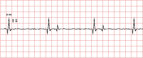
A 33-year-old woman has noted increasing difficulty with her secretarial work as the day progresses, making more mistakes at her computer keyboard and seemingly unable to keep her eyes open to see the computer monitor. She tries getting more sleep each night, but this does not improve her condition. She exhibits progressive weakness and fatigue over the next several months. When seen by her physician, she is noted to have more proximal muscular weakness, particularly with repetitive motion. Deep tendon reflexes are normal. No neurologic deficits are noted. Cranial nerves II - XII are intact. She has normal mental function. There is no muscle pain. Her range of motion in her extremities is normal. No joint pain is present.
The results of electromyography are shown here:
A week later she develops respiratory distress and goes to the emergency room. She requires intubation. The blood bank is consulted to arrange for emergent plasmapheresis.
-
- Which of the following laboratory test findings is most likely to be present in this patient?
- A. Positive serology for Lyme disease
- B. Positive acetylcholine receptor antibody
- C. Elevated creatine kinase
- D. Increased cortisol
- E. Monoclonal gammopathy
|
Further History:
The patient has a chest CT scan performed, and there is a 6 cm mass noted in the anterior mediastinum.
-
- What is this mass most likely to be?
- A. Nodular sclerosis Hodgkin's disease
- B. Small cell anaplastic carcinoma
- C. Tuberculous lymphadenitis
- D. Thymoma
- E. Abscess
|
Further History:
Following surgery for removal of the anterior mediastinal mass, she still remains weak for several years.
-
- Which of the following therapies following surgery is most likely to be effective for this patient over the next several years to treat the underlying problem?
- A. Corticosteroids
- B. Antibiotics
- C. Plasmapheresis
- D. Chemotherapy
- E. Physical therapy
|
|
CASE 2
(Click here to go to the answers)
Clinical History
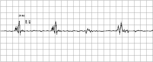
A 5-year-old boy is noted by his parents to be less active than other children. The child has recently had increasing difficulty getting up the stairs to his room. The parents are concerned, because the mother's sister had a child with similar findings who died at age 19. The family's primary care physician examines the boy and finds no deformities or abnormalities. The boy's height and weight are normal for age. Deep tendon reflexes are normal. However, the child has only 4/5 motor strength in upper and lower extremities. Despite prominence of the boy's calves, he has difficulty getting up from a sitting position on the floor.
The results of electromyography are shown here:
-
-
-
-
-
- Which of the following laboratory test findings is most likely to be present?
- A. High antinuclear antibody titer
- B. Positive acetylcholine receptor antibody
- C. Increased sweat chloride
- D. Peripheral blood eosinophilia
- E. Markedly elevated creatine kinase
|
Further History:
A gastrocnemius biopsy is performed and the images are reviewed.
-
- What is most likely to be seen on light microscopy?
- A. Marked variation in myofiber size and marked fibrosis between fibers
- B. Acute inflammation with necrosis and abscess formation
- C. Grouped atrophy of myofibers without inflammation or fibrosis
- D. Extensive lymphocytic infiltrates between myofibers
- E. Infiltration of myofibers by hyperchromatic and pleomorphic cells
|
Further History:
An immunohistochemical stain is performed that demonstrates absence of a protein on the muscle fibers.
-
- What is this muscle fiber protein?
- A. Fibrillin
- B. Spectrin
- C. Beta myosin
- D. Dystrophin
- E. Acetylcholine
|
- What was the probable immediate cause of death in the boy's 19-year-old cousin?
- A. Cardiomyopathy
- B. Acute renal failure
- C. Bronchopneumonia
- D. Cerebral edema with herniation
- E. Malignant lymphoma
|
Further history:
The boy's step-father has a brother who had the onset of mild limb weakness at age 20. This weakness was mainly proximal. A biopsy showed a decrease, but not absence, of the same muscle fiber protein by immunohistochemical staining.
-
- What disease is the boy's step-uncle most likely to have?
- A. The same disease
- B. Becker muscular dystrophy
- C. Werdnig-Hoffman disease
- D. Myophosphorylase deficiency
- E. Amyotrophic lateral sclerosis
|
|
CASE 3
(Click here to go to the answers)
Clinical History
While travelling through a small rural village, a couple of university students spot a street vendor selling sausages. They are hungry, and the price is cheap. One student comments, "I wonder where those came from. There isn't any supermarket here, and I haven't seen a butcher shop." Hunger is the deciding factor. The sausages taste good, even though they're cold. They move on to their next destination. However, two days later, they both develop abdominal pain, nausea, and diarrhea. One of them notes a mild fever. These symptoms subside, but a week later both students have muscle pain, weakness, and fever.
-
-
- Which of the following laboratory test findings is most characteristic for this disease?
- A. Elevated antistreptolysin O titer
- B. Lactic acidosis
- C. Urine positive for myoglobin
- D. Increased troponin I
- E. Peripheral blood eosinophilia
|
- Which of the following food handling practices would most likely have prevented the occurrence of this disease?
- A. Thorough washing
- B. Serving with alcoholic beverage
- C. Thorough cooking
- D. Removing the sausage casing
- E. Thorough chewing
|
- Which of the following animals is most likely to be the intermediate host for this process?
- A. Snail
- B. Chicken
- C. Horse
- D. Catfish
- E. Pig
|
|
CASE 4
(Click here to go to the answers)
Clinical History
A 30-year-old woman has an ultrasound performed in the 18th week of pregnancy. The ultrasound reveals no anomalies of major organs, but fetal movement is decreased. The baby is born at term. The umbilical cord is short. The baby is noted to be weak and floppy. Muscle stretch reflexes are absent. However, the baby is alert and responds to stimuli. The baby's weakness rapidly worsens. Death occurs at 8 months of age from respiratory failure with pneumonia.
-
-
- What histopathologic finding is most likely to be seen in skeletal muscle?
- A. Marked variation in fiber size along with marked fibrosis between muscle fibers
- B. Acute inflammation with necrosis and abscess formation
- C. Grouped atrophy of muscle fibers without inflammation or fibrosis
- D. Extensive lymphocytic infiltrates between muscle fibers
- E. Marked glycogen deposition within muscle fibers
|
- What is the most appropriate statement to make to the mother regarding this condition?
- A. You should have supplemented your diet with folate during pregnancy
- B. A vaccination for poliovirus should prevent this from happening again
- C. This is a sporadic condition not likely to occur again
- D. In subsequent pregnancies, there is a 25% recurrence risk
- E. A karyotype on fetal cells can help identify this condition
|
- What disease process with similar symmetrical and progressive muscular weakness has an onset in adults?
- A. Amyotrophic lateral sclerosis
- B. McArdle's disease
- C. Becker muscular dystrophy
- D. Myasthenia gravis
- E. Pompe's disease
|
|
CASE 5
(Click here to go to the answers)
Clinical History
A 15-year-old adolescent has been bothered by painful muscle cramps during physical education classes at school. This has been occurring over the past couple of months. His coach just tells him, "Work it off with a few more laps around the track." The boy believes that there is a more serious problem, because his urine is darker following these episodes. The boy's family physician refers him to a neurologist. The neurologist determines that electromyography is normal at rest.
-
-
- Which of the following laboratory test findings in the patient's blood is most likely to be present?
- A. Increased D-dimer
- B. No rise in lactate after exercise
- C. Positive antinuclear antibody test
- D. Increased troponin I
- E. Normal serum creatine kinase
|
- Which of the following histopathologic findings is most likely to be seen in a skeletal muscle biopsy?
- A. Lack of staining for dystrophin
- B. Grouped atrophy
- C. Diminished size and number of type II fibers
- D. PAS positive deposits
- E. Infiltration by T cells
|
- Which of the following organs is most likely to be affected by this condition?
- A. Liver
- B. Brain
- C. Kidney
- D. Heart
- E. Adrenal
|
- Which of the following long-term outcomes of this patient's disease is most likely to occur?
- A. Inability to perform competitive sports
- B. Neurologic deterioration
- C. Hepatic cirrhosis
- D. Congestive heart failure
- E. Chronic renal failure
|
Explain the biochemistry of this disease.
|
CASE 6
(Click here to go to the answers)
Clinical History
A 24-year-old woman presented with a slowly progressive muscle weakness. She first noted difficulty in getting out of a chair some months ago and now has difficulty climbing stairs. The weakness is not made significantly worse by repetitive activity, nor does it go away with rest. She also noted some muscle pain associated with this illness. On physical examination, she has moderate proximal muscle weakness. Laboratory findings include a serum creatine kinase of 3534 U/L. An electromyogram demonstrates a combination of myopathic features and fibrillation potentials.
-
-
- Which of the following serologic test findings is most likely to be present in this case?
- A. Anti-histidyl tRNA synthetase
- B. Anti-double stranded DNA
- C. Anti-parietal cell antibody
- D. Anti-DNA topoisomerase I
- E. Anti-ribonucleoprotein
|
Further history:
A month later she has a faint violet coloration to the skin of her eyelids and cheeks, but without significant erythema, and it is not made worse by sun exposure. She continues to have muscular weakness. A muscle biopsy is performed. By light microscopy with H&E staining, there is focal inflammation with lymphocytes, along with rare scattered plasma cells and neutrophils. Some of the inflammation is endomysial. Some of the muscle fibers show degeneration with necrosis. A few muscle fibers appear to be regenerating.
-
- Which of the following pathologic mechanisms most likely accounts for these histologic findings?
- A. Antigen-antibody complexes
- B. Infection by Staphylococcus aureus
- C. Lack of a cell membrane stabilizing protein
- D. Wallerian degeneration
- E. Antibody-mediated cytotoxicity
|
- Which of the following organs is most likely to be affected by this condition?
- A. Liver
- B. Brain
- C. Kidney
- D. Heart
- E. Adrenal
|
- In adults with this condition, which of the following underlying diseases is more likely to occur?
- A. Membranoproliferative glomerulonephritis
- B. Atherosclerotic heart disease
- C. Gastric adenocarcinoma
- D. Hyperparathyroidism
- E. Pulmonary emphysema
|
Further history:
Her condition does not improve. How is consent for her treatment obtained when she no longer has capacity?.
|
CASE 7
(Click here to go to the answers)
Clinical History
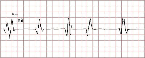
A 34-year-old woman has experienced increasing difficulty with activities that involve movement over the past year. She now finds it hard to climb even one flight of stairs. She cannot lift a chair and carry it across a room. She cannot walk more than 300 m.
On examination, she has no muscle tenderness, but large proximal muscle groups exhibit decreased tone. She has 3/5 motor strength in upper and lower extremities for proximal muscles such as quadriceps and biceps femoris, gluteal muscles, deltoids, and biceps brachii. There is 4/5 motor strength for distal muscle groups in hands and feet.
Laboratory studies show Hgb 14 g/dL, creatinine 0.8 mg/dL, and creatine kinase 155 U/L. The results of electromyography are shown here:
-
-
- What is seen on light microscopy?
- A. Marked variation in myofiber size and marked fibrosis between fibers
- B. Acute inflammation with necrosis and abscess formation
- C. Grouped atrophy of myofibers without inflammation or fibrosis
- D. Extensive lymphocytic infiltrates between myofibers
- E. Infiltration of myofibers by hyperchromatic and pleomorphic cells
|
Further history:
Six months later she develops difficulty speaking and swallowing. Explain the pathogenesis of her disease and correlate with the findings above.
|
|
 Return to the
Laboratory Menu.
Return to the
Laboratory Menu.
