Clinical History:
A 5-year-old boy is noted by his parents to be less active than other children. The child has recently had increasing difficulty getting up the stairs to his room. The parents are concerned, because the mother's sister had a child with similar findings who died at age 19. The family's primary care physician examines the boy and finds no deformities or abnormalities. The boy's height and weight are normal for age. Deep tendon reflexes are normal. However, the child has only 4/5 motor strength in upper and lower extremities. Despite prominence of the boy's calves, he has difficulty getting up from a sitting position on the floor.
The images show muscle fiber atrophy with interstitial fibrosis. The muscle of the patient with Duchenne muscular dystrophy exhibits no staining for dystrophin. The pedigree is typical for an X-linked pattern of inheritance (female carriers indicated by a dot in the circle). Female carriers may pass the trait onto their sons, who are affected, or to their daughters, who become carriers. Males can only pass the trait to daughters. Some woman carriers may have mild muscular weakness, depending upon random X-inactivation.
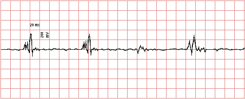
The results of electromyography are shown here:

This EMG shows a "myopathic" pattern with discharges characterized by summation of small, short duration, polyphasic motor unit action potentials, typically with four or more phases.
-
- Which of the following laboratory test findings is most likely to be present?
- A. High antinuclear antibody titer
- B. Positive acetylcholine receptor antibody
- C. Increased sweat chloride
- D. Peripheral blood eosinophilia
- E. Markedly elevated creatine kinase
|
Answer: E The findings at this age are most suggestive of Duchenne muscular dystrophy, particularly in view of the family history. This is a myopathic process, so the CK should be increased.
Further History:
A gastrocnemius biopsy is performed and the images are reviewed.
-
- What is most likely to be seen on light microscopy?
- A. Marked variation in myofiber size and marked fibrosis between fibers
- B. Acute inflammation with necrosis and abscess formation
- C. Grouped atrophy of myofibers without inflammation or fibrosis
- D. Extensive lymphocytic infiltrates between myofibers
- E. Infiltration of myofibers by hyperchromatic and pleomorphic cells
|
Answer: A These findings are characteristic for Duchenne muscular dystrophy.
Further History:
An immunohistochemical stain is performed that demonstrates absence of a protein on the muscle fibers.
-
- What is this muscle fiber protein?
- A. Fibrillin
- B. Spectrin
- C. Beta myosin
- D. Dystrophin
- E. Acetylcholine
|
Answer: D Duchenne muscular dystrophy is an X-linked disorder that is due to a defective gene on the short arm of the X chromosome leading to an inability to produce dystrophin, which is part of the membrane skeleton. The destabilization of the muscle fibers from the lack of dystrophin leads to the eventual loss of the fibers. In half of cases, there is a woman carrier in the family (who may have an increased creatine kinase but not clinical symptoms of the disease); in the remaining half, a spontaneous new mutation is present, since this is a large gene susceptible to mutations.
-
- What was the probable immediate cause of death in the boy's 19-year-old cousin?
- A. Cardiomyopathy
- B. Acute renal failure
- C. Bronchopneumonia
- D. Cerebral edema with herniation
- E. Malignant lymphoma
|
Answer: C Persons who are disabled with chronic neuromuscular diseases are at risk for aspiration and/or pneumonia, and this is a common cause for death.
Further history:
The boy's step-father has a brother who had the onset of mild limb weakness at age 20. This weakness was mainly proximal. A biopsy showed a decrease, but not absence, of the same protein by immunohistochemical staining.
-
- What disease is the boy's step-uncle most likely to have?
- A. The same disease
- B. Becker muscular dystrophy
- C. Werdnig-Hoffman disease
- D. Myophosphorylase deficiency
- E. Amyotrophic lateral sclerosis
|
Answer: B There is reduced (but not absent) dystrophin with Becker muscular dystrophy, a milder dystrophy with onset of weakness in later childhood to young adulthood with a slower and more variable rate of progression.
|
|


