Criminalistics Laboratory Methods
Surgical pathology description of bullets
Each bullet keeps a diary in its own way of where it has been and
what it has done. Now that you understand the function of a bullet, many of
these changes become easy to interpret. The bullet base will contain irregular
dimples marking the pressure delivered there in its acceleration. the bullet
sides will bear the markings of the barrel interior rifling. These spiral
lines, or striae, contain the micrscopic imperfections of the gun from which it
was fired and can be as specific as a fingerprint. The bullet nose carries
information about the target, and recognizing these may give a clue to the
injury rendered. Remember in measuring bullets to determine the type of cartridge used
that the actual bullet diameter, even of non-deformed bullets, is not the same
as the name of the cartridge. Most names have a historic basis and have little
to do with any real physical measurements: a .30-06 was named for a .30 caliber
cartridge developed in 1906; the handgun cartridges called .357 magnum, .38
special, and 9 mm parabellum have essentially the same .357 inch actual
diameter. Therefore, use caution in opinions regarding the type of weapon or
cartridge used based upon examination of bullets. The best surgical pathology description would give dimensions as
measured (use vernier calipers for best results), shape, and appearance of
surface. Photography will be valuable. Expansion of a semi-wadcutter hollowpoint bullet increases the frontal
area and blunts the shape. The degree to which this happens depends upon the
texture of the tissue impacted, the velocity at impact, and the softness of the
bullet (usually quite constant). With the exceptions of lung and bone, tissue
densities are relatively constant. Velocity is the most important factor. No change in shape occurs until impact velocity achieves about 800 fps.
Between 800 and 1000 fps a slight flattening of the bullet nose can be
expected. Over 1000 fps real expansion starts to occur and by 1200 fps the nose
is turned over to form a mushroom shape. An interesting artefact of impacts
around 1000 fps is the tendency of the copper jacket to be shed from the lead.
The jacket stops in the subcutaneous tissue and the bullet will continue to
penetrate. This accounts for fragments of copper (with rifling marks) commonly
seen as surgical specimens. At velocities approaching 1500 fps the bullet is
transformed into a rounded ball of lead and copper. The above results are
uniformly valid only in artificial media (such as ordnance gelatin) but
correlate with human tissue. Examples follow on the next page: The soft exposed lead nose on non-full metal jacketed bullets can be
imprinted with anything that is penetrated by the bullet. Wood, glass, fabric,
plastic, or tissue may leave marks as well as fragments on the bullet tip. Bone
struck by bullets may not only fragment the bone, but also split the bullet.
Lead round nose bullets can penetrate deeply and strike bone at relatively high
velocity and can be cleanly cut in half or shaved vertically. Full metal
jacketed round nose bullets are less affected, but are often irregularly
flattened upon striking bone. Bullets that come to rest in soft tissue without
striking bone are often intact. Intermediate targets, such as glass, wood, clothing, or even paper, may
influence the path, shape, and fragmentation of projectiles. Such factors must
be taken into account in the recovery of evidence. (Stahl et al, 1979) Even
tempered glass, which shatters and fragments easily on impact, may deflect
handgun bullets (low velocity) significantly. High velocity, jacketed bullets
will be deflected much less. (Thornton, 1986) Flattening of shotgun pellets may not necessarily indicate a close
range contact with a target, as the pellets may be deformed on firing. Recently
developed shells use plastic packing materials and plastic capping to diminish
deformation. (DeMuth et al, 1978) Even pellets of air guns may show characteristic striae (Cohle et al,
1987). Silencers used over the muzzle of a gun are often misaligned and can
produce characteristic striae. (Menzies et al, 1981)
Examination of whole bullets in crime laboratory
If a bullet is recovered from the scene or from the body, it may
be compared to bullets obtained by test-firing the suspected weapon. Test
firing is done using similar ammunition. Bullets are marked on the nose at the
12 o'clock barrel position (called "index", "witness", or "reference"
marks). Consecutive test bullets are then fired into a water tank, recovered,
and juxtaposed with a comparison microscope to compare test bullets with the
recovered evidence. Index marks help to align test bullets to determine
reproducibility of markings. Photographs should be taken (a ruler or coin can
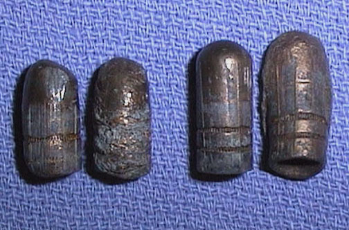
be used to give scale and alignment). Comparison of bullets involves "class" and "individual" characteristics. These characteristics are based upon "striae" left on the bullet as it passes through the barrel. Class refers to the type of caliber and rifling. Rifling pattern may
turn to the right or left, with a given rate of twist. The number and depth of
grooves can vary also. Some newer guns use "polygonal" rifling resembling the reversed image of a twisted square rod. A particular type of gun (.38 Smith and Wesson, or 9 mm Glock) will impart these class characteristics. Individual characteristics are used to try and determine if a specific
gun (say one of many 9 mm Glocks) was used. These individual characteristics
are based upon burrs or imperfections in the barrel, particularly the muzzle,
that impart specific markings, or striae, to fired bullets. If such markings
are present, they may lead to a "determinative" identification. In
general, smaller caliber weapons (.22) yield fewer reproducible characteristics
in fired bullets than weapons of larger caliber (.45). In the image below, two sets of bullets of the same class are roughly compared to indicate how difficult this can be when bullet deformation is present. Patterns of Striae on Bullets
A system has also been described for identification of jacketed
sporting rifle bullets using twelve parameters:
- Identification number
- Manufacturer
- Weight
- Diameter
- Cartridge
- Base design
- Length of bearing surface
- Color
- Shape
- Location and description of crimping cannelure
- Location and description of other cannelures
- Miscellaneous notes.
Such parameters may aid in narrowing the search for suspected weapons or ammunition. (Booker, 1980) There are three results of comparison identification. Test fired and
recovered bullets can: (1) be related to the same weapon; (2) be unrelated to
the same weapon; (3) not be compared with this type of examination.
Conclusions should not be based upon probabilities in test firing. In many situations, however, the hospital pathologist as medical
examiner will not be involved with test firing. The hospital pathology
department may receive bullets or bullet fragments from patients. Such evidence
should be clearly identified, with a "chain of custody" followed. The
pathologist will dictate a report and release the evidence back to the
authorities.
Examination of bullet fragments or bullet composition
In many cases, recovered bullets will be too deformed for
comparison studies. A method has been described for differentiation of bullets
by spark source mass spectrometry (SSMS). This method makes use of the fact
that the "lead" of bullets actually may contain up to 26 common
elements, of which 12 can be used for differentiation. One of the commonest of
these is antimony (1 to 2%) Unfortunately, the study also found that bullets
within a box or lot do not have uniform composition, but there may be two or
three distinct groups of bullets within a box. Such a study may have limited
usefulness in some cases. (Haney and Gallagher, 1975) When analysis of the bullet lead is necessary, but a copper jacket is
present, the copper may be most efficiently removed, without contamination of
the lead, by use of concentrated nitric acid. (Izak-Biran et al, 1980) Detection of the type of bullet (jacketed or not) may be done by a
dithiooxamide (rubeanic acid) test. This test detects copper and nickel, which
may be components of jacketed ammunition, on the target. The rubeanic acid
forms a dark green precipitate in the presence of copper, pink or blue with
nickel, and brown with cobalt. Blood and other materials on the target produced
false negatives. (Lekstrom and Koons, 1986) Bullet particles may also be detected in bone fragments from skeletal
remains when no soft tissues remain. After determining that radiopaque
particles are present, surfaces of the bone fragments containing the particles
can be exposed by cutting. The surfaces can then be analyzed by SEM-EDA and by
electron probe microanalysis to identify lead (Pb) and antimony (Sb). The
electron probe technique aids in differentiating antimony from abundant calcium
of bone. (Simmelink et al, 1981) Detection of bullet lead has also been carried
out with proton-induced X-ray emission (PIXE) analysis, even in a victim buried
for several years (Fischbeck et al, 1986). Even if an exit wound is present, a search for bullet fragments or
jacket material should be done, with radiographs if necessary. A new type of
ammunition, Winchester Western Silvertip, may pose a problem, as its aluminum
jacket is only faintly radiopaque (Conradi, 1982).
|
 Return to the Firearms Tutorial menu.
 Proceed to Examination of Gunshot Residue.