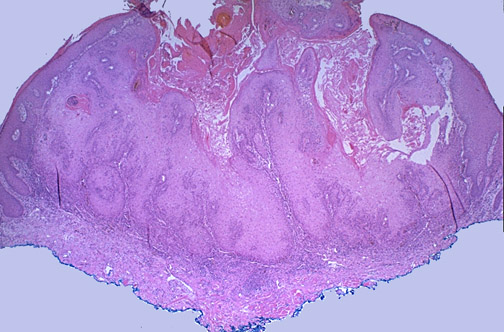
The low power view of an excised keratoacanthoma seen here shows the cup shaped pattern of growth, with a central area with keratin debris surrounded by proliferating squamous epithelium (the blue ink is used to mark the margins of the excision).