
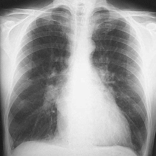
| The posterior-anterior (PA) chest radiograph seen here demonstrates marked pulmonary congestion and edema throughout all lung fields. The pulmonary veins are distended near the hilum. The left heart border is prominent due to left atrial enlargement. This patient had mitral stenosis. |


 |