
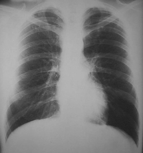
| This PA chest radiograph reveals extensive granulomatous disease of both lungs. Note the focal calcifications that are typical for healed tuberculosis. Other small white calcifications are scattered, mainly in mid to upper lung fields, and more on the right, as seen here. There can be reactivation or reinfection to produce this pattern for secondary tuberculosis. |


 |