
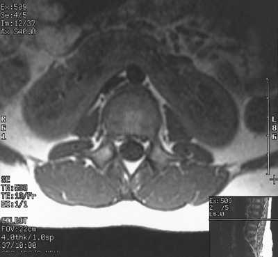
| This abdominal MRI scan with contrast reveals a horseshoe kidney bridging across the midline. In this view the contrast media is excreted into and highlights the renal pelves of the fused kidney. The path of the ureters over the top of the bridge between the fused lower poles is potentially a point of obstruction. |


 |