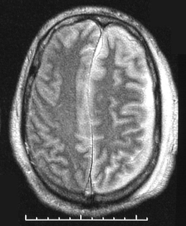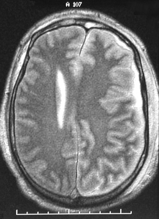
| The T2 weighted MRI scans in axial view above and below show bright fluid in the subdural region on the left, representing a subdural empyema. Note also the effacement of the left ventricle with midline shift due to the brain swelling from the inflammation. This may represent hematogenous spread of infection, or a prior subdural hematoma may become infected. Both events are not common. |
|
|