|
A 23-year-old man suddenly collapses to the ground while practicing tai-kwan-do. He is taken to the emergency department. Vital signs show T 36.9 C, P 88/min, RR 30/min, and BP 110/70 mm Hg. Auscultation of the chest reveals absent breath sounds on the right. There is tympany on the right. His heart rate is regular with no murmurs. No other significant findings are noted.
An arterial blood gas shows pO2 55 mm Hg, pCO2 30 mm Hg, pH 7.45, and HCO3 28 meq/L. After administration of 100% oxygen by nasal canula at 2 L/min, a repeat blood gas shows pO2 55 mm Hg, pCO2 30 mm Hg, pH 7.45, and HCO3 27 meq/L
A chest radiograph is taken.
Questions:
2.1 What was the sudden event?
Pneumothorax
2.2 Name conditions that may have predisposed to this event.
Paraseptal (localized) emphysema can have rupture of a peripheral bulla. An inhaled foreign body with atelectasis could produce a similar effect. Chest trauma with penetrating injury or rib fracture could do the same.
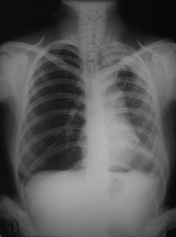
A chest radiograph confirms the right pneumothorax, with a mediastinal shift to the left.
Tympany is the phenomenon of a low-pitched sound coming from a hollow structure, such as a drum, the stomach, or a collapsed lung. As the beer keg empties, it becomes more tympanitic, and you percuss the side of the keg to determine the level.
2.3 Explain the blood gas findings.
Administration of increased oxygen will have no effect, since this is a ventilation defect.
There is a V/Q mismatch.
Normally the rate of inspiration is about 14 breaths per minute. The tidal volume of a normal adult is about 500 mL. Thus, the minute ventilation is about 7 L, but anatomic dead space reduces this by 2L, so the alveolar ventilation is 5L/minute.
Ventilation and perfusion (the V/Q ratio) are closely matched in normal lung, with a value of 0.8 (since there is a physiologic V/Q mismatch from greater ventilation in upper lung fields and greater perfusion in lower lung fields).
If there is no ventilation but the lung is still perfused, then the V/Q ratio is 0, and the non-ventilated area is essentially a shunt. Blood leaving this non-ventilated area retains the blood gas parameters of venous blood entering the lung. Examples of this include airway obstruction, atelectasis, and alveolar filling processes (alveolar exudates with pneumonia, pulmonary edema). A low PO2 will not correct with administration of oxygen.
If there is ventilation, but no perfusion, then the V/Q ratio is high and the non-perfused area acts as dead space, though what little blood exits has blood gas parameters approaching that of the alveolar air. Examples of this include pulmonary arterial obstruction with a thromboembolus, vasculitis, and diseases reducing the pulmonary capillary bed such as obstructive and restrictive lung diseases. Giving oxygen will improve the PO2.
| 
