- Why is this marriage tanking?
What we have here is a failure to communicate. Putting aside the issue of "selective hearing loss" from psychosocial causes, not pathologic causes, and that counseling is in order, then pathologic conditions need to be considered.
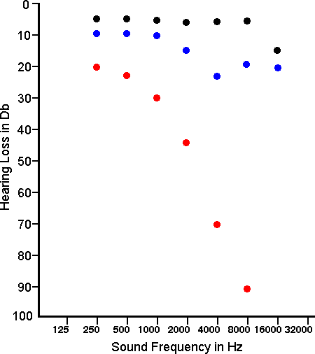
- How do you interpret the audiogram (3 patients represented)?
The black dots represent near normal hearing in a young adult. If the blue dots represent a young adult, then there is premature hearing loss, particularly for high-pitched sounds, worst around 3000 to 4000 Hz, typical for noise damage. If the red dots represent the patient (husband) in this case, there could be presbycusis.
The theoretical maximal range of human hearing is between 20 and 20,000 Hz. Normal human speech is in the range of 250 to 6000 Hz. The auditory canal enhances sounds in the 2000 to 5000 Hz range.
- What diseases could lead to these findings? How are they distinguished on physical examination?
Physical examination can help to distinguish whether the process is unilateral or bilateral. Testing for air conduction and bone conduction can be done. The range of hearing loss can be tested by audiometry. Etiologies for a conductive form of hearing loss include:
Otitis Media: middle ear infections are relatively common, and most of these clear up without seqaelae. The hearing loss, if any, is temporary. One may see a red eardrum or fluid bulging behind the eardrum.
Perforated eardrum (tympanic membrane): this may complicate a severe infection or could result from trauma.
Cholesteatoma: not a true neoplasm but a cystic structure lined by squamous epithelium that can form following repeated middle ear infections or mastoiditis. It can enlarge over time and destroy structures in its pathway.
Ossicular Chain Discontinuity: trauma with fracture of the temporal bone may also involve the ossicles.
Otosclerosisis: this problem is an autosomal dominant condition that affects perhaps 10% of Caucasians, but in 90% of cases it is mild and does not lead to significant hearing loss. This condition is chacterized by growth of irregular woven bone that can cause ankylosis of the ossicles, typically at the footplate of the stapes anchoring it to the oval window, to produce "bone conduction" hearing loss. Surgery can be done to mobilize the ossicles or put in a replacement prosthesis.
Ear wax: cerumen (ear wax) can build up in the external auditory canal. It is a simple matter to wash it out.
Etiologies for a sensorineural form of hearing loss include:
Meniere's Disease: there is formation of endolymphatic hydrops, and a major symptom is tinnitus (ringing of the ears). Obstruction of the endolymphatic duct will result in progressive increase in the volume and biochemical degradation of endolymph. The sacculus dilates and frequently contacts the stapes footplate. This is followed by a more diffuse distortion of the membranous labyrinth. Over time endolymphatic hydrops leads to varying degrees of degeneration of the organ of Corti, stria vascularis, hair cells, and cochlear neurons.
Labyrinthitis: Infection producing inflammation may reach the inner ear via the middle ear, the meninges, or rarely by hematogenous dissemination. This inflammation primarily affect the semicircular canals to produce vertigo but may involve adjacent cochlear structures. Labyrinthitis ossificans is the pathologic ossification of spaces in the membranous labyrinth that occurs in response to a destructive process. Labyrinthitis ossificans has etiologies that include trauma, otosclerosis, and viral infection, but the most common cause is bacterial infection of the inner ear resulting in suppurative labyrinthitis Inflammation usually occurs in the perilymphatic spaces, typically sparing the endolymphatic space. As early as 2 weeks after the onset of meningitis, fibrosis is present, followed by new bone formation, which may be seen within 2 months. While ossification is accompanied by degenerative changes in the stria vascularis and organ of Corti, the auditory nerve fibers are retained to a variable degree.
[The vestibular system has six semicircular canals that sense angular (rotational) head movements and two utricles and saccules to sense linear head movements and the acceleration of gravity. The semicircular canals connect, via bipolar neurons, to the vestibular nuclei (VN), which project to the oculomotor nuclei to complete the vestibulooculomotor reflex. This reflex is responsible for stabilizing images on the retina during head movements.]
Meningitis: some persons experience hearing loss following a bout of bacterial meningitis. It is one of the more important causes for hearing loss in children. Hearing loss during meningitis occurs due to the spread of bacteria or bacterial toxins through the internal auditory canal or cochlear aqueduct, causing a suppurative labyrinthitis or perineuritis of the eighth nerve, or both
Sudden sensorineural hearing loss: this condition may follow viral infection, but the etiology is often not known. About 2/3 of cases resolve spontaneously.
Presbycusis (presbyacusis): degeneration of hair cells and ganglion cells can be associated with aging, but a more specific etiology should be sought.
Noise damage: the external hair cells of the organ of Corti in the cochlea are very sensitive structures. Repeated exposure to loud noises (high decibel range), including jet aircraft, vehicles without mufflers, and rock music concerts, leads to damage and loss of the hair cells. The loss is predominantly in the range of high-pitched sound. Once the hair cells are gone, they are gone. The damage is permanent.
Congenital: such as from rubella infection).
Ototoxicity: from drugs such as aminoglycosides and aspirin.
Mitochondrial mutations: several mutations in mitochondrial genes, both inherited and acquired, have been implicated in some cases of presbycusis. Reactive oxygen metabolites (ROM) are known products of oxidative metabolism and are continuously generated in vivo. The ROM are extremely reactive and cause extensive DNA, cellular, and tissue damage. Specific deletions within the mitochondrial DNA (mtDNA) occur with increasing frequency in age from chronic exposure to ROM and can contribute to presbyacusis. When enough mtDNA damage accrues, the cell becomes bioenergetically deficient. This mechanism is the basis of the mitochondrial clock theory of aging, also known as the membrane hypothesis of aging.
Schwannoma: the eighth cranial nerve can be damaged by inflammation and by neoplasia (such as a schwannoma). The neoplasm can be removed, but the hearing loss on the affected side is permanent in many cases. Hearing is preserved in some. An auditory brainstem implant can be tried in some cases with deafness following surgery.
- How are they distinguished on physical examination?
Use the tuning fork to help distinguish conductive from sensorineural hearing loss.
Weber Test (for lateralization): Place the base (not the prongs) of the lightly vibrating tuning fork firmly on the midline of patient's head or on the mid-forehead. For normal hearing, sound is heard equally in both ears. With conductive hearing loss, sound lateralizes to the bad ear, but to the good ear with sensorineural hearing loss.
Rinne Test (compares air conduction with bone conduction): Place the base of the lightly vibrating tuning fork on the mastoid bone behind the ear and on level with the ear canal (this is testing bone conduction). When the patient can no longer hear the sound, quickly place the "U" of the fork close to the ear canal and ascertain whether the sound can still be heard (this is testing air conduction). Normally, the sound is heard longer through air than through bone (Air Conduction > Bone Conduction). Bone conduction is equal to or greater than air conduction of there is a conductive hearing loss. Air conduction lasts longer with sensorineural hearing loss.
Patients with conductive hearing loss have normal speech discrimination, but patients with sensorineural hearing loss have poor speech discrimination.
Severity of hearing loss: