X-ray crystallography research illustration
Functional materials research illustration
X-ray diffraction pattern
Structural disorder illustration

Exploring the structure of functional materials

We develop and apply X-ray crystallographic methods to probe material structures. We investigate structural features across different length scales to obtain crystallographic, structural disorder and microstructural information. We study materials under electrical, thermal and, in the future, mechanical stimuli. We currently focus on piezoelectrics and ferroelectrics but are open to exploring other material systems in which the relationship between structure and properties is not yet understood.

Current research projects

Diagram of crystal domains and high-resolution diffraction

Investigation of multidomain crystals using high-resolution X-ray diffraction

A domain is a finite region of a crystal in which a physically meaningful order parameter is uniform. Domains and domain walls may mediate physical properties such as giant piezoelectricity and shape-memory effects. We develop methods for investigating domains using X-ray and neutron scattering, identifying domains from Bragg-peak splitting and tracking their response to external fields.

Time-resolved X-ray single-crystal diffraction setup

Development of time-resolved single-crystal X-ray diffractometry

X-ray diffraction is a leading tool for investigating material structures. Understanding the structure–property relationship requires measuring how a structure responds to external conditions. This response may involve changes in chemical bond lengths, macroscopic strain, or variations in the microstructure and domain pattern. For example, applying an electric field to a piezoelectric material induces macroscopic deformation that may arise from intrinsic structural changes or domain-wall motion. We develop data-acquisition systems that synchronize X-ray diffraction measurements with the application of external stimuli, allowing us to probe a device in operation.

Illustration of crystallographic cleavage planes

Crystallography of cleavage

Cleavage describes the tendency of a crystal to break along specific lattice planes. Information about cleavage in a given crystal structure is essential for investigating key mechanical properties such as fracture toughness, plasticity and strength. Although the cleavage planes of simple crystals, such as silicon, are well known, this information is not generally available for arbitrary crystal structures.
We are developing a universal algorithm to automatically inspect crystal structures and predict their likely cleavage planes. The algorithm is being implemented as a MATLAB program. The project is carried out in collaboration with Prof Dov Sherman and his brittle fracture laboratory.

Group Members

Principal investigator

PhD students

Ido Biran

Ido Biran

PhD student

Research interests: High-resolution X-ray diffraction, Phase transitions

Elena Pelegova

Elena Pelegova

PhD student

Research interests: Birefringence microscopy, Ferroelectric materials, Imaging of domains

Master’s students

Oren Daniel

Oren Daniel

MSc student

Lab engineer

Active visitors

Alumni & former visitors (7 people)

News

2023 (4 stories)
Visiting the Australian Synchrotron

Visiting the Australian Synchrotron

It was a pleasure to visit the Australian Synchrotron today. During our visit, we presented our research and toured the beamlines. Last but not least, we explored Melbourne’s southeastern suburbs and the area around Monash University.

Beamtime at the Swiss-Norwegian Beamlines

Beamtime at the Swiss-Norwegian Beamlines

We are performing an experiment at the mighty Swiss-Norwegian Beamlines at the European Synchrotron in Grenoble. All eyes and ears are on Dmitry Chernyshov.

On our way to Grenoble

On our way to Grenoble

We are on our way to Grenoble, France! A week-long synchrotron experiment with plenty of X-ray photons, data and food lies ahead. We are already starting with the last item on that list at the airport. Our long journey takes us via Istanbul and Geneva.

Powder diffraction experiments at SESAME

Powder diffraction experiments at SESAME

We are at SESAME in Allan, Jordan, today. After spending a day exploring Amman, we are preparing for a multi-temperature powder diffraction experiment at the MS beamline. Meanwhile, we are enjoying the cool weather, beautiful views and plenty of food.

2021 (2 stories)
A glimpse inside the lab

A glimpse inside the lab

Not really news, but a nice view of the lab! The red light is on and data collection is under way. Nobel Prize? Maybe tomorrow!

Welcome, Yair Dror

Welcome, Yair Dror

We welcome Yair Dror, our new laboratory engineer. He will spend half of his time with us and the other half with Dr Maxim Sokol’s ceramics laboratory. We wish Yair a smooth start and many successful and enjoyable years of work. And, of course, we wish him good health during these challenging times.

2020 (9 stories)
A new analyzer for time-resolved diffraction

A new analyzer for time-resolved diffraction

The wait is over: the multichannel analyzer for time-resolved X-ray diffraction experiments with a point detector has arrived. It was developed by our former colleagues at the University of Siegen. It is great to see an old friend.

A closer look at our X-ray beam

A closer look at our X-ray beam

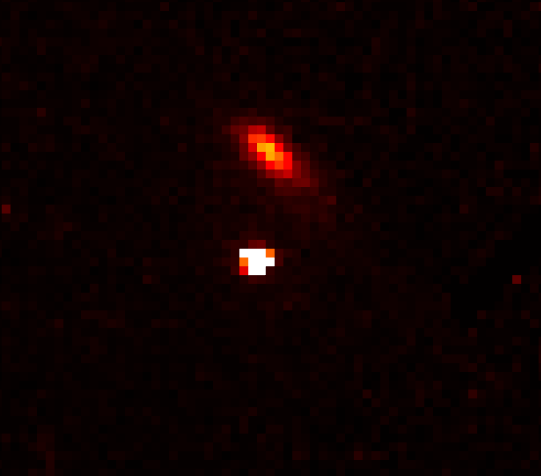
Here is the beam as it emerges from the X-ray microfocus source and parabolic X-ray mirror. The spots on the right and left contain the Kα and Kβ lines, respectively. The Kβ line can be eliminated with a pinhole, while Kα1 will be selected by the monochromator. More details will appear in an upcoming paper.

Reciprocal space reflected in the detector

Reciprocal space reflected in the detector

The “reality” of reciprocal space: an X-ray diffraction pattern reflected from the surface of the detector. Nothing unusual, just a beautiful image.

Studying crystals under electric fields

Studying crystals under electric fields

Our time-resolved X-ray diffraction measurements under an external electric field continue. Does this material have a chance to become the new “star” of piezoelectricity? If so, why? We will get the answer soon.

First electric-field experiments in the lab

First electric-field experiments in the lab

Another milestone to celebrate in the lab: our first laboratory-based experiments on crystals under an electric field have begun. This time, we are performing test measurements of the piezoelectric coefficient of a quartz single crystal. The PILATUS detector is synchronized with the applied electric field using this methodology.

New paper accepted in Applied Physics Letters

New paper accepted in Applied Physics Letters

Our new paper has been accepted for publication in Applied Physics Letters. The work resulted from an earlier beamtime at the ID22 beamline of the European Synchrotron. It was led by our major collaborators Dr Christopher Fancher from ORNL and Prof Jacob Jones from NCSU, USA.

Crystallography of cleavage: our first paper

Crystallography of cleavage: our first paper

We are happy to announce that our first article on crystallography of cleavage in functional materials has been published in the latest issue of Acta Materialia. This work resulted from our collaboration with Prof Dov Sherman and his brittle fracture laboratory.

Research during the lockdown

Research during the lockdown

During the COVID-19 outbreak, our lives are dominated by self-isolation, Zoom meetings and online teaching. The laboratory is closed and experiments are suspended. This period may nevertheless be useful for analytical research, data analysis and revisiting older, unpublished algorithms.

2019 (20 stories)
New photographs of our laboratory

New photographs of our laboratory

A few colorful photographs of our laboratory are now available here. Special thanks to Limor Ben Moshe for the excellent photography.

Mapping reciprocal space with CrysAlisPro

Mapping reciprocal space with CrysAlisPro

We can now use CrysAlisPro software to search for and index reflections in single-crystal diffraction data. The reciprocal-space reconstruction from a Na0.5Bi0.5TiO3 crystal shows that even diffuse X-ray scattering is detectable.

Presenting at ECM32 in Vienna

Presenting at ECM32 in Vienna

We are on our way to Vienna for ECM32, the European Crystallographic Meeting. Dr S. Gorfman will give a talk, and U. Vaknin will present a poster. We are looking forward to it!

Late nights on campus

Late nights on campus

Sometimes it is necessary to work late, especially when another important conference is approaching. At least the night views across the Tel Aviv University campus are relaxing and beautiful.

Diffractometer installation complete

Diffractometer installation complete

The installation is now complete! Several intensive weeks of work with the wonderful Youli Li from Forvis Technologies are over, and our X-ray diffractometer is almost ready for its research challenges.

Support from the US-Israel Binational Science Foundation

Support from the US-Israel Binational Science Foundation

We have received a positive response from the US-Israel Binational Science Foundation. For three years beginning in October 2019, the foundation will support our collaborative research with Dr Igor Levin from the National Institute of Standards and Technology. The work will focus on elucidating the local structure and disorder in functional ferroelectric oxides.

Exploring high scattering angles

Exploring high scattering angles

The real crystallography begins when the X-ray detector can move to a high scattering angle relative to the incident beam. Our custom-built instrument is certainly able to do that.

Aligning our first crystal

Aligning our first crystal

Every X-ray crystallographer knows the excitement of mounting a new sample on a diffractometer. It is even more exciting when the instrument has just been installed and this is its first alignment.

First image from the PILATUS detector

First image from the PILATUS detector

The PILATUS 1M X-ray area detector is switched on, and the honor of producing its first image goes to a cosmic ray! Cosmic particles are everywhere, and there is no way to hide from them. During this 100-second exposure, the detector captured several of their characteristic streaks.

Assembling the diffractometer

Assembling the diffractometer

At the end of the first installation day, the instrument components have been lifted onto the optical table. Setting up the communication channels, routing cables through the ceiling, and switching on the source and detectors come next.

Installing the goniometer

Installing the goniometer

The installation moves forward! The goniometer has been lifted onto the table. There is still a long way to go: we must set up the X-ray source, monochromator chamber, detectors and communication cables.

Diffractometer installation begins

Diffractometer installation begins

This is a truly historic moment: installation of the X-ray diffractometer begins! The four-circle goniometer, a key part of the instrument, enters the lab.

Our diffractometer arrives

Our diffractometer arrives

It has finally arrived! Two boxes weighing a total of 500 kg have completed a 15,000 km journey, landed in Israel and reached Tel Aviv University. Our diffractometer will be installed next week!

Wishing Min success in his new position

Wishing Min success in his new position

Sometimes things change very quickly in academia. We congratulate Min on his new position in the United States and wish him every success. Fortunately, we will be able to continue our fruitful collaboration.

Setting up the electronics bench

Setting up the electronics bench

The electronics bench is taking shape. The oscilloscope, multimeter and high-frequency voltage amplifier are learning how to work together. “To share or not to share the earth—that is the question.”

Working through ideas at the whiteboard

Working through ideas at the whiteboard

A whiteboard is, and has always been, an important instrument in any research laboratory. Feeling stuck? Our advice is to step back, take a deep breath and come to the board. It will always help in one way or another. Here, Min is developing an electronic circuit for in situ diffraction studies of ferroelectrics.

A fast X-ray detector arrives

A fast X-ray detector arrives

After a long wait, a fast LaBr3 scintillation counter has arrived at our lab from FMB Oxford. It will soon be integrated into the diffractometer to detect X-ray photons in time-resolved X-ray diffraction experiments.

A custom germanium monochromator

A custom germanium monochromator

This germanium crystal was manufactured for the monochromator in our X-ray diffractometer by Integratds in Slovakia. Its unusual configuration features asymmetric cuts with two different asymmetry angles. The monochromator is optimized for beam transmission.

Welcome, Dr Hyeokmin Choe

Welcome, Dr Hyeokmin Choe

We are happy to welcome Dr Hyeokmin Choe (Min). He joins our group as a postdoctoral researcher. Min is originally from Seoul, South Korea, but has lived in the United States for the past year and a half. He completed his PhD at the University of Siegen. We will certainly benefit from his experience in designing, performing and analyzing in situ X-ray diffraction experiments.

2018 (3 stories)
Experiments at ESRF beamline ID31

Experiments at ESRF beamline ID31

We performed another experiment at the European Synchrotron Radiation Facility. This time, we worked at beamline ID31 in collaboration with Tony Schenk from the Luxembourg Institute of Science and Technology.

Israel Science Foundation funding

Israel Science Foundation funding

For the next four years, our research will be supported by an Israel Science Foundation grant: “Fine structure and polarization rotation in ferroelectric perovskites.”

Our research on the journal cover

Our research on the journal cover

Our latest paper is featured on the cover page of Journal of Applied Crystallography. This paper demonstrates how synchrotron diffraction experiments with an area detector can explain the origin of enhanced piezoelectricity in ferroelectric single crystals. The experiments were performed at the Swiss-Norwegian Beam Lines at the ESRF.

Latest publications

2026 (5 publications)

Measurements and scaling of X-ray total scattering from single crystals

S. Gorfman, M. Eremenko, V. Krayzman, A. Bosak, P. Y. Zavalij, I. Levin

Journal of Applied Crystallography, 59, 1224–1237 (2026)

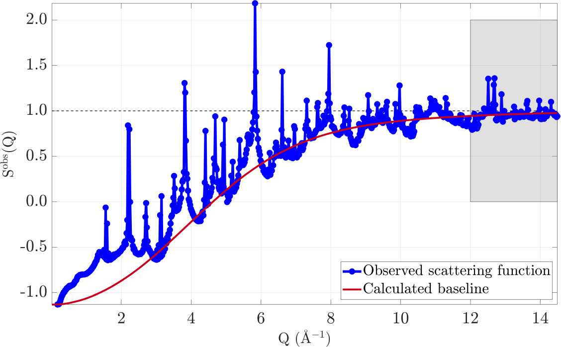
Scattering functions used to scale X-ray total-scattering data from single crystals

Epitaxial FePd thin films on SrTiO3(001): Kinetics of single-variant L10 ordering and solid-state dewetting

H. K. Dara, S. Vergara, A. Azulay, M. Uemoto, T. Ono, S. Gorfman, I. Goldfarb, H. Naganuma, A. Kohn

Acta Materialia, 314, 122333 (2026)

Research figure: Epitaxial FePd thin films on SrTiO3(001)

Asymmetry of the Ferroelectric Phase Transition in BaTiO3

A. Hershkovitz, E. Hemaprabha, R. Mandal, P. Kavle, J. Tanus, M. Barzilay, C.-C. Lin, D. Spirito, S. Gorfman, B. Wang, I. J. Villar-García, N. Domingo, L.-Q. Chen, L. W. Martin, Y. Ivry

Advanced Materials, 38(25), e16507 (2026)

Research figure: Asymmetry of the Ferroelectric Phase Transition in BaTiO3

Twinning relationships in coexisting cubic and tetragonal phases of ferroelectrics

S. Gorfman, I. Biran, N. Zhang, Z.-G. Ye

Acta Materialia, 304, 121762 (2026)

Research figure: Twinning relationships in coexisting cubic and tetragonal phases of ferroelectrics

Identification and characterization of monoclinic domains from three-dimensional single-crystal diffraction patterns in ferroelectric perovskites

G. Zhang, V. Dyadkin, D. Chernyshov, Z.-G. Ye, S. Gorfman, N. Zhang

Acta Materialia, 303, 121727 (2026)

Research figure: Identification and characterization of monoclinic domains from three-dimensional single-crystal diffraction patterns in ferroelectric perovskites
2025 (2 publications)

Emergent topological polarization textures in relaxor ferroelectrics

M. Eremenko, V. Krayzman, S. Gorfman, A. Bosak, H. Y. Playford, P. A. Chater, B. Ravel, W. J. Laws, F. Ye, A. Minelli, B.-X. Wang, Z.-G. Ye, M. G. Tucker, I. Levin

Nature Communications, 16, 7531 (2025)

Research figure: Emergent topological polarization textures in relaxor ferroelectrics

Structural transformations in the (1−x)Na0.5Bi0.5TiO3−xBaTiO3 solid solution under high pressure: Comparison between x below and above the morphotropic phase boundary

C. Rösche, T. Malcherek, T. Boffa Ballaran, C. Paulmann, R. J. Angel, S. Gorfman, B. Mihailova

Physical Review B, 112, 184102 (2025)

Research figure: Structural transformations in the (1−x)Na0.5Bi0.5TiO3−xBaTiO3 solid solution under high pressure: Comparison between x below and above the morphotropic phase boundary
2024 (4 publications)

The high-pressure structure of (1-x) NaBi0.5Ti0.5O3 -x BaTiO3 at the morphotropic phase boundary

C. Rösche, T. B. Ballaran, T. Malcherek, C. Paulmann, R. J. Angel, S. Gorfman, B. Mihailova

Scientific Reports, 14, 18799 (2024)

Research figure: The high-pressure structure of (1-x) NaBi0.5Ti0.5O3 -x BaTiO3 at the morphotropic phase boundary

Lattice match between coexisting cubic and tetragonal phases in PMN-PT at the phase transition

I. Biran, A. Bosak, Z.-G. Ye, I. Levin, S. Gorfman

Applied Physics Letters, 124, 242901 (2024)

Research figure: Lattice match between coexisting cubic and tetragonal phases in PMN-PT at the phase transition

Permissible domain walls in monoclinic ferroelectrics. Part II. The case of MC phases

I. Biran, S. Gorfman

Acta Crystallographica Section A, 80, 293–304 (2024)

Research figure: Permissible domain walls in monoclinic ferroelectrics. Part II. The case of MC phases

Permissible domain walls in monoclinic MAB ferroelectrics phases

I. Biran, S. Gorfman

Acta Crystallographica Section A, 80, 112–128 (2024)

Research figure: Permissible domain walls in monoclinic MAB ferroelectrics phases
2022 (3 publications)

“Forbidden” Polarisation and Extraordinary Piezoelectric Effect in Organometallic Lead Halide Perovskites

M. Vasiljevic, M. Kollár, D. Spirito, L. Riemer, L. Forró, E. Horváth, S. Gorfman, D. Damjanovic

Advanced Functional Materials, 32, 2204898 (2022)

Research figure: “Forbidden” Polarisation and Extraordinary Piezoelectric Effect in Organometallic Lead Halide Perovskites

Identification of a coherent twin relationship from high-resolution reciprocal-space maps

S. Gorfman, D. Spirito, G. Zhang, C. Deltlefs, N. Zhang

Acta Crystallographica Section A, 78, 158–171 (2022)

Research figure: Identification of a coherent twin relationship from high-resolution reciprocal-space maps

Induced giant piezoelectricity in centrosymmetric oxides

D.-S. Park, M. Hadad, L. M. Riemer, R. Ignatans, D. Spirito, V. Esposito, V. Tileli, N. Gauquelin, D. Chezganov, D. Jannis, J. Verbeeck, S. Gorfman, N. Pryds, P. Muralt, D. Damjanovic

Science, 375(6581), 653–657 (2022)

Research figure: Induced giant piezoelectricity in centrosymmetric oxides
2021 (4 publications)

Geometrical prediction of cleavage planes in crystal structures

U. Vaknin, D. Sherman, S. Gorfman

IUCrJ, 8(5), 793–804 (2021)

Research figure: Geometrical prediction of cleavage planes in crystal structures

Multipurpose diffractometer for in situ X-ray crystallography of functional materials

S. Gorfman, D. Spirito, N. Cohen, P. Siffalovic, P. Nadazdy, Y. Li

Journal of Applied Crystallography, 54(3), 914–923 (2021)

Research figure: Multipurpose diffractometer for in situ X-ray crystallography of functional materials

Mean inner potential of graphite measured by electron holography: Probing charge distribution and orbital diamagnetic susceptibility

A. Auslender, G. Levi, V. Ezersky, S. Gorfman, O. Dieguez, A. Kohn

Carbon, 179, 288–298 (2021)

Research figure: Mean inner potential of graphite measured by electron holography: Probing charge distribution and orbital diamagnetic susceptibility

Quantitative mapping of nanotwin variants in the bulk

J. Schultheiss, L. Porz, L. K. Venkataraman, M. Hoefling, C. Yildirim, P. Cook, C. Detlefs, S. Gorfman, J. Roedel, H. Simons

Scripta Materialia, 199, 113878 (2021)

Research figure: Quantitative mapping of nanotwin variants in the bulk
2020 (6 publications)

Algorithms for target transformations of lattice basis vectors

S. Gorfman

Acta Crystallographica Section A, 76(6), 713–718 (2020)

Research figure: Algorithms for target transformations of lattice basis vectors

Electrically driven transient and permanent phase transformations in highly strained epitaxial BiFeO3 thin films

H. J. Lee, J. Kim, S. H. Hwang, H. Choe, S. Gorfman, Y.-J. Heo, S. Y. Lee, P.-E. Janolin, I. Kornev, T. U. Schuelli, C. Richter, J.-H. Lee, U. Pietsch, C.-H. Yang, J. Y. Jo

APL Materials, 8, 101110 (2020)

Research figure: Electrically driven transient and permanent phase transformations in highly strained epitaxial BiFeO3 thin films

Impact of polishing on crystallinity and static performance of Cd1-xZnxTe

A. Brovko, A. Adelberg, L. Chernyak, S. Gorfman, A. Ruzin

Nuclear Instruments and Methods in Physics Research A, 984, 164568 (2020)

Research figure: Impact of polishing on crystallinity and static performance of Cd1-xZnxTe

Effect of Alloying BaTiO3 with BiZn0.5Ti0.5O3 on Polarization Reversal

C. M. Fancher, H. Choe, S. Gorfman, H. Simons, C. C. Chung, M. Ziolkowski, S. Prasertpalichat, D. P. Cann, J. L. Jones

Applied Physics Letters, 117, 042907 (2020)

Research figure: Effect of Alloying BaTiO3 with BiZn0.5Ti0.5O3 on Polarization Reversal

New method to measure domain-wall motion contribution to piezoelectricity: the case of PbZr0.65Ti0.35O3 ferroelectric

S. Gorfman, H. Choe, G. Zhang, N. Zhang, H. Yokota, A. M. Glazer, Y. Xie, V. Dyadkin, D. Chernyshov and Z.-G. Ye

Journal of Applied Crystallography, 53(4), 1039–1050 (2020)

Research figure: New method to measure domain-wall motion contribution to piezoelectricity: the case of PbZr0.65Ti0.35O3 ferroelectric

Cleavage and surface energies of LiNbO3

Y. Hirsh, S. Gorfman, D. Sherman

Acta Materialia, 192, 338–349 (2020)

Research figure: Cleavage and surface energies of LiNbO3
2019 (2 publications)

Intrinsic Ferroelectricity in Charge-Ordered Magnetite

M. Angst, S. Adiga, S. Gorfman, M. Ziolkowski, J. Strempfer, C. Grams, M. Pietsch, J. Hemberger

Crystals, 9(11), 546 (2019)

Research figure: Intrinsic Ferroelectricity in Charge-Ordered Magnetite

Local-scale structural response of (1-x)Na0.5Bi0.5TiO3-xBaTiO3 to external electric fields

G. D. L. Flor, S. Gorfman, B. Mihailova

Applied Physics Letters, 114(4), 042901 (2019)

Research figure: Local-scale structural response of (1-x)Na0.5Bi0.5TiO3-xBaTiO3 to external electric fields
2018 (5 publications)

Probing the intrinsic and extrinsic origins of piezoelectricity in lead zirconate titanate single crystals

N. Zhang, S. Gorfman, H. Choe, T. Vergentev, V. Dyadkin, H. Yokota, D. Chernyshov, B. Wang, A. M. Glazer, W. Ren, Z.-G. Ye

Journal of Applied Crystallography, 51(5), 1396–1403 (2018)

Research figure: Probing the intrinsic and extrinsic origins of piezoelectricity in lead zirconate titanate single crystals

Ferroelectric domain wall dynamics characterized with X-ray photon correlation spectroscopy

S. Gorfman, A. A. Bokov, A. Davtyan, M. Reiser, Y. Xie, Z.-G. Ye, A.V. Zozulya, M. Sprung, U. Pietsch, C. Gutt

Proceedings of the National Academy of Sciences, 115(29), E6680–E6689 (2018)

Research figure: Ferroelectric domain wall dynamics characterized with X-ray photon correlation spectroscopy

Monoclinic distortion, polarization rotation and piezoelectricity in the ferroelectric Na0.5Bi0.5TiO3

H. Choe, J. Bieker, N. Zhang, A. M. Glazer, P. A. Thomas, S. Gorfman

IUCrJ, 5, 417–427 (2018)

Research figure: Monoclinic distortion, polarization rotation and piezoelectricity in the ferroelectric Na0.5Bi0.5TiO3

Picometer polar atomic displacements in strontium titanate determined by resonant X-ray diffraction

C. Richter, M. Zschornak, D. Novikov, E. Mehner, M. Nentwich, J. Hanzig, S. Gorfman, D. C. Meyer

Nature Communications, 9(1), 178 (2018)

Research figure: Picometer polar atomic displacements in strontium titanate determined by resonant X-ray diffraction

Local-scale structures across the morphotropic phase boundary in PbZr1-xTixO3

N. Zhang, H. Yokota, A. M. Glazer, D. A. Keen, S. Gorfman, P. A. Thomas, W. Ren, Z.-G. Ye

IUCrJ, 5(1), 73–81 (2018)

Research figure: Local-scale structures across the morphotropic phase boundary in PbZr1-xTixO3
2017 (2 publications)

Structural transformations in (1-x)Na0.5Bi0.5TiO3 - x BaTiO3 single crystals studied by Raman spectroscopy

G. D. L. Flor, T. Malcherek, S. Gorfman, B. Mihailova

Physical Review B, 96(21), 214102 (2017)

Research figure: Structural transformations in (1-x)Na0.5Bi0.5TiO3 - x BaTiO3 single crystals studied by Raman spectroscopy

Time and frequency-dependence of the electric field-induced phase transition in BaTiO3-BiZn1/2Ti1/2O3

T. Iamsasri, G. Esteves, H. Choe, M. Vogt, S. Prasertpalichart, D. P. Cann, S. Gorfman, J. L. Jones

Journal of Applied Physics, 122, 064104 (2017)

Research figure: Time and frequency-dependence of the electric field-induced phase transition in BaTiO3-BiZn1/2Ti1/2O3

Funding

Israel Science Foundation

Enabling accurate X-ray structural analysis and domain reconstruction in ferroelectric PbZr1-xTixO3 crystals

Project duration: October 2023 – October 2027

Research illustration for the ISF structural-analysis project

Joint NSFC-ISF (China–Israel) Research Grant

Phase coexistence and strain-free boundaries in single crystals of functional ferroic materials

Project duration: October 2021 – October 2024

Research illustration for the joint NSFC-ISF project

DFG: Deutsche Forschungsgemeinschaft

The mechanisms of enhanced electromechanical coupling in uniaxial ferroelectrics

Project partner: Dr Carsten Richter, Leibniz Institute for Crystal Growth

Project duration: October 2023 – October 2026

Research illustration for the DFG electromechanical-coupling project

BSF: US-Israel Binational Science Foundation

Local structure mechanisms of electromechanical coupling in oxide ferroelectrics

Project partner: Dr Igor Levin, National Institute of Standards and Technology, Gaithersburg, USA

Project duration: October 2019 – October 2026

Research illustration for the BSF local-structure project

Israel Science Foundation

Fine structure, polarization rotation and low-symmetry phases in ferroelectric perovskites

Project duration: October 2018 – October 2023

Research illustration for the ISF ferroelectric-perovskites project

Contact us

Wolfson Building for Mechanical Engineering