 |
 |
||
Structural Model of Penile Erection |
 |
||
|
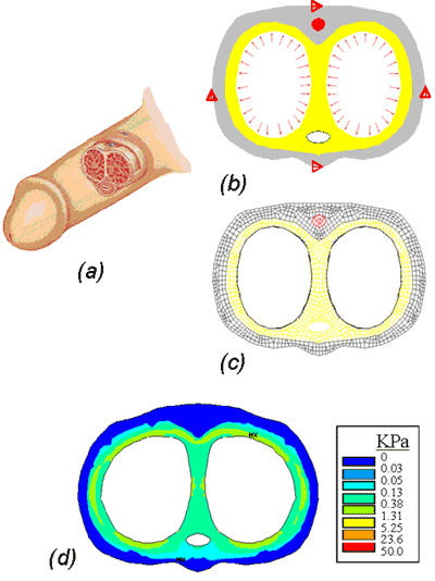
Amit Gefen1, Chen1,2, David Elad1 The boundary conditions included 4 constraints on the lateral and dorsal-plantar aspects of the penis, allowing its expansion as a result of inflation by an equivalent erectile pressure Ρe = Ρa- σcc. The erectile pressure, Ρe, reflected the resistance stress σcc of the spongy corpus cavernosa tissue to inflation pressure Ρa = 100 mmHg caused by arterial blood flow into the cavities of the penis. As the penis becomes erect, blood is supplied to the corpus cavernosa until the full erection corporal volume reaches VE which is the total corporal capacity (TCC). When blood drains and the penis becomes flaccid, the corporal volume reduces up to VF = 35% TCC. Assuming that the corpus cavernosal tissue is unstressed at VF, the characteristic stretch ratio λmax from flaccid to full erection is given by the generally accepted relationship λ= (VE/VF)1/3 and equals 1.42. It is further assumed that the mechanical characteristics of the corpus cavernosal tissue is similar to those of the lung parenchyma, hence, σcc=7 KPa at λmax=1.42. The penile soft tissues were assumed to be homogenous, isotropic, and linear elastic materials. The simulation of stress distribution in the normal penis indicated that most of the load bearing during inflation is carried by the dorsal part of the tunica albuginea, where stresses are in the range of 5-30 KPa (Fig. 1b). Since this site contains several nerves, it is most vulnerable to intensified mechanical stresses. The skin appeared to bear a negligible load. Inflation of the neutral, elliptical cross-sectional shape of the cavernosum during erection yielded a more circular corporal profile as well as lateral expansion of the cross-sectional shape of the penis (Fig. 1). This modeling approach allows simulation of stress distributions in various pathologic conditions of the penis (e.g., diabetes, Peyronie’s disease) by altering the geometry and material properties of its components.
References
|
|||