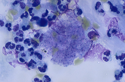
With Giemsa staining of a smear of fluid from bronchoalveolar lavage (BAL), the small dot-like
intracystic bodies
(not the cyst wall) of P. carinii are visible within the exudate.