


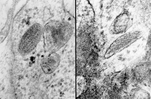
| Sometimes a melanoma will not be well-differentiated enough to show the typical melanin pigmentation by light microscopy. By electron microscopy (EM), it may be possible to prove the neoplasm is a melanoma if premelanosomes are seen. Two examples of premelanosomes are shown here as oval structures with a faint barred pattern. |
|
|