 Return to the radiologic techniques menu.
Return to the radiologic techniques menu.
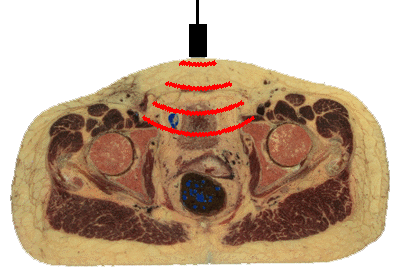
Ultrasound (US) uses high-frequency sound waves for scanning body regions. Ultrasonography is a non-invasive technique that does not use x-rays, so it is safe for any patient. US is commonly utilized for prenatal diagnosis for this reason. To perform US, a transducer generates a focused beam of sound waves that is directed toward the body region to be studied. The sound beam is oriented in a specific plane, such as sagittal (longitudinal) or axial (transverse). The transducer is moved across the external body surface while the image appears on a monitor.
The sound beam srikes body structures that echo (reflect) the sound waves to varying degrees--tissues have different acoustic impedances. The reflected sound waves are detected by the transducer and converted to a series of electrical impulses that is computed to form an image viewed on a screen. Selected views can be captured on film. However, the real advantage of US is the ability to move the transducer in real time and see what is happening. Movement can be detected, such as the beating of a fetal heart. A video can be made of the images to record the movement. Ultrasonographic images are not sharp, but "fuzzy" and require skill in interpretation. A single image is not as useful as a real time exam.
The image displayed on the monitor depends upon the echogenicity of the structures beneath the beam. Fluids are echolucent, so the bladder and the gallbladder appear dark. Solid organs and soft tissues are echogenic. Structures with calcium are very echogenic. A gallstone appears bright with US. Bone is so dense to sound waves that it cannot be evaluated. This puts the brain and spinal cord "off limits" for US examination. US is most suited to study of heart with its valves (which move), gallbladder, vessels (to check for thrombi and flow), and internal genitalia.
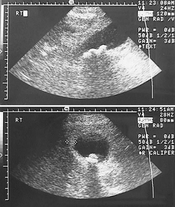
Shown below is an ultrasound of the upper abdomen in a transverse plane that demonstrates the echolucent lumen of the gallbladder with a bright gallstone present. Note how the stone creates an echo "shadow" below it.
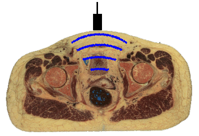
Movement can be further enhanced by "doppler" ultrasonography in which flow away from the transducer is converted to a signal that appears blue, while flow toward the transducer appears reddish. This can be useful in recording flow in arteries and veins.