Return to the radiologic techniques menu.
Return to the radiologic techniques menu.
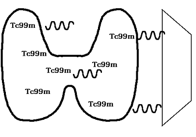
Nuclear medicine makes use of radioisotopes injected intravenously that are taken up more selectively by certain tissues. Over a period of minutes to hours, the radioactive decay of the isotope is detected with a gamma camera. The radioisotopes chosen have a short half-life, making them safe, and they are typically excreted into the urine.
One of the most common radioisotopes utilized is technetium 99m (Tc99m), which is easily generated just prior to use. The Tc99m can be attached to substances that make uptake more selective. For example, a combination of Tc99m with micro-aggregated albumin will be trapped in peripheral pulmonary capillaries, allowing scanning of the lungs to be accomplished.
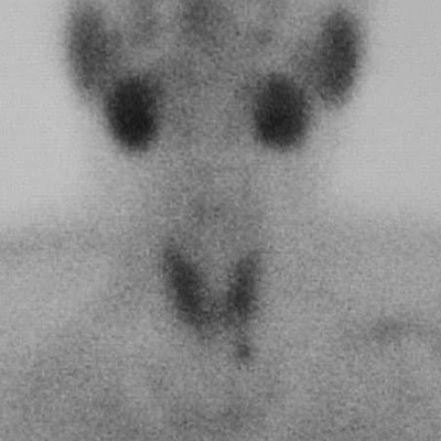
In the example below, the uptake is into the thyroid gland, with emission of gamma rays that are detected by the camera at the right.
A scan that demonstrates the thyroid, but highlights a parathyroid adenoma, is seen below.
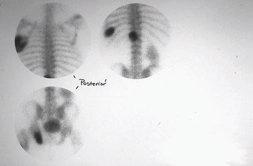
One of the most useful nuclear medicine scans is a bone scan. The whole skeleton can be imaged. Areas of increased bone turnover have more radioisotopic uptake and are "hot spots" that appear darker on the exposed film. In the scan below, there are multiple "hot spots" representing metastases. Can you guess the primary site? (One kidney is absent.)