 Return to the radiologic techniques menu.
Return to the radiologic techniques menu.
Computerized axial tomography, or computed tomography (CT) is a way of using x-rays to create cross sectional views of the body. A thin beam of x-rays is moved around the patient while electronic detectors opposite the beam produce an electrical output. The output varies according to the tissues traversed by the x-ray beam. Each axial slice can be divided into a grid of pixels, or picture elements representing a small part of the overall image. (Alternatively, the pixels are called voxels because they represent unit volumes.) Each individual pixel (voxel) has its own density.
The CT scanner rotates the x-ray beam around the patient to create each slice.
The information obtained from the beam detectors is analyzed by a computer that computes the brightness of each pixel. The resulting image can be displayed on a monitor or printed onto x-ray film.
The brightness of a pixel in a CT image is based upon the x-ray absorption of the tissue at that point. The range of brightness depends upon the absorption values assigned to the tissues. By convention, water has a value of 0 Hounsfield units (Geoffrey Hounsfield was one of the inventors of CT imaging). Denser structures have a higher number and air has the lowest (negative) number. thus, bone is bright white and air is black. This attenuation can be altered to make the structures visualized darker or brighter, depending upon the desired view. These attenuation "windows" are picked for the best view of the body region or structures that need to be evaluated. Thus, there are windows for lung, soft tissue, brain, bone, and liver.
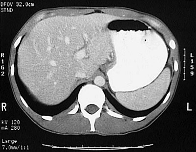
A CT scan, therefore, consists of a series of axial (transverse) slices through the body taken at intervals ranging from 0.5 to 1 cm. By convention, a slice is viewed as though you were looking up from the patient's feet toward the head, so the CT image has the patient's left side on your right. The patient is typically positioned prone in the scanner, so the top of the CT image is the patient's ventral aspect (front). (Radiologists look up, pathologists look down.) If you can't remember, look for the "L" and the "R" on the image.
CT images tend to be quite sharp, but artefacts in the CT image can occur. The most common is the scattering of x-rays into a starburst pattern from very dense materials such as bone or metallic objects (earrings, surgical clips, etc). Movement by the patient, such as occurs with breathing, can blur the images. However, with high speed CT scanners this is generally not a problem.
Conventional CT scanners produce a series of axial images over a 10 to 20 minute period per body region. Fast CT scanners move the x-ray beam around the patient in a continuous spiral motion so that a complete scan of a body region may take less than 90 seconds. Such a high-speed CT scan keeps movement artefacts to a minimum.
Patients are often given contrast material to drink, typically dilute water soluble iodinated contrast or dilute barium sulfate suspension, in the case of abdominal CT scans, to help outline the gastrointestinal tract, particularly the small intestine. Contrast agents may be given intravenously to help highlight vessels. A small number of patients may have an adverse reaction to contrast. Oral contrast should be avoided with bowel obstruction. If the patient's renal function is not adequate, intravenous contrast should not be given.