
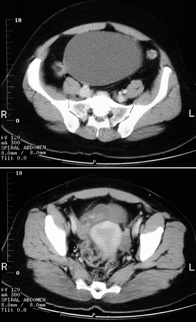
| A large cystic mass is seen filling much of the pelvis in the upper panel of this pelvic CT scan. The unilocular mass has a thin wall and is fluid-filled. The inferior margin of the mass can be seen to attach to the right ovary next to the bladder, where the wall is somewhat thickened and irregular. This is a benign ovarian cystadenoma. |


 |