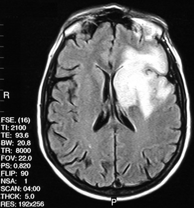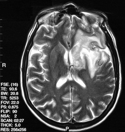
| The axial T2 weighted FSE MRI scan above and the FLAIR MRI scan below both demonstrate an area of enhancement involving primarily the left frontal white matter. There was also extension to basal ganglia, thalamus, and portions of the temporal and parietal lobes in this case of progressive multifocal leukoencephalopathy (PML). Note the relative sparing of the overlying cortex seen best in the FLAIR scan below. |


 |