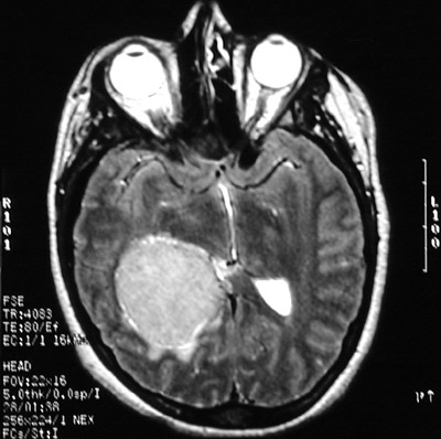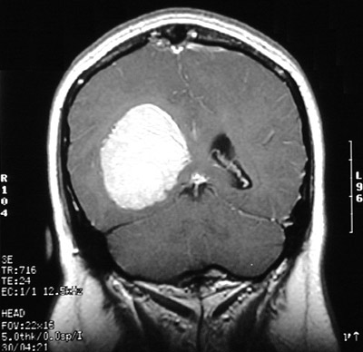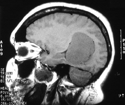
| The T1 weighted MRI scan in axial view above demonstrates another less common location for a meningioma, an intraventricular meningioma. In this location, a circumscribed mass could also be an ependymoma. Below can be seen the same right lateral ventricular mass with contrast enhancement in T2 weighted axial view and T1 weighted coronal view. |
|
|