


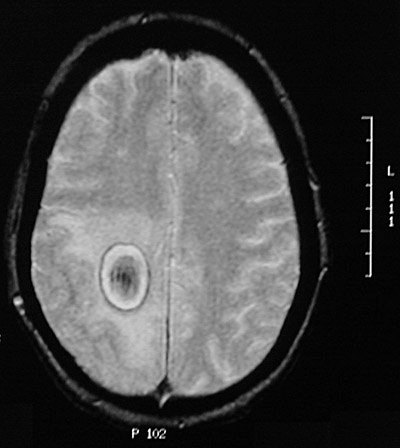
| This is an organizing hematoma in the parietal lobe seen with a T2 weighted MRI scan in axial view. It is distinguished from an abscess because the center is not bright from liquid contents. It is distinguished from a neoplasm by the dark rim representing hemosiderin deposition and the dark center with the blood products. A necrotic center would be white on a T2 weighted scan, more typical for a neoplasm with central necrosis or an abscess. There is some surrounding edema. |
|
|