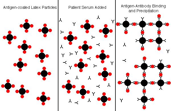
Latex Agglutination
The latex agglutination (LA) assay is a simple and inexpensive method for determining if antibodies to a variety of viral agents are present.
In the example below, the latex particles are coated with antigen, and the presence of specific antibody in the patient serum will result in agglutination of the particles.
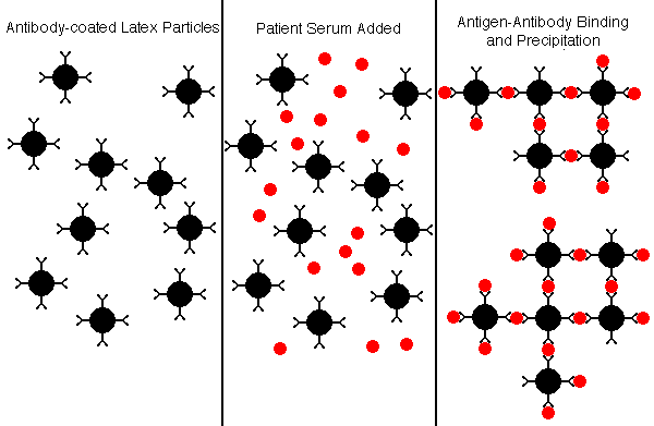
Alternatively, in the example below, the latex particles are coated with antibody, and the presence of specific antigen in the patient serum will result in agglutination of the particles.
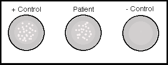
A rapid latex particle agglutination test is often performed using reagents and samples on a card, and the agglutination reaction is read from the card after a short incubation time of a minute or two.
Complement Fixation
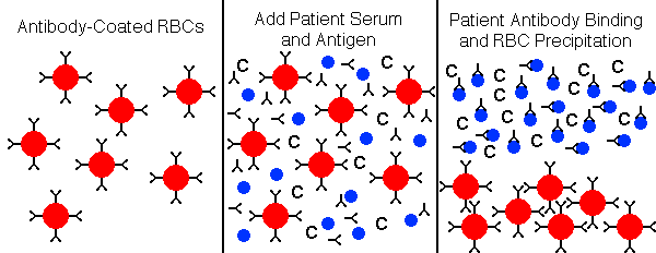
Complement fixation (CF) tests have largely been replaced by enzyme immunoassays which have greater sensitivity. In the example shown below, patient serum, which normally contains complement components, is added along with antigen (such as viral antigen) to the tube or well. If no patient antibody is present to the antigen, then the antigen will attach to the RBCs, and the antigen-antibody complex will fix complement that lyses the RBCs, leading to hemolysis.
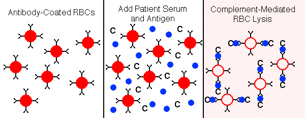
In the example below, the patient serum contains specific antibody to the antigen, forming soluble antigen-antibody complexes, while the RBCs precipitate to the bottom of the tube or well.
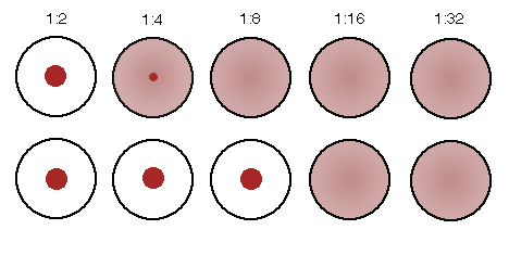
The patient serum to be tested is often serially diluted, in order to provide an estimate of the strength of the antibody titer. In the example below, the top row represents the "acute" titer of patient antibody (the initial antibody response, or persistent antibody from past exposure to the antigen). The bottom row represents the "convalescent" titer from a sample drawn 2 weeks later, when the secondary antibody response occurred. The four-fold increase in titer from acute to convalescent suggests that a recent encounter with the antigen (such as an acute viral infection) occurred. It is now more common to measure IgM and IgG specific antibody on a single sample to determine if an acute response has occurred.
Hemagglutination Inhibition
Hemagglutination inhibition (HI) is a technique that is based upon a surface feature of some viruses which have hemagglutinin spikes on their surfaces that can attach to receptors on RBCs and cause agglutination (clumping) of the RBCs. If there are specific antibodies to the viral hemagglutinins, then the antibody binding prevents the viruses from attaching to the RBCs, and agglutination of the RBCs is inhibited.
The test is performed by adding serial dilutions of patient serum to a series of wells that contain RBCs and viral antigen. When sufficient patient antibody is present to bind to viral antigen, then agglutination is inhibited and the RBCs precipitate to the bottom of the well, forming a button. When there is no patient antibody present, then the virus will cause agglutination to form a lattice or web that keeps the RBCs in suspension and prevents them from sinking to the bottom of the well.
In the example below, serial dilutions of patient sera are added to wells containing suspensions of RBCs and viral antigen, such as influenza A. Rows A and B represent acute and convalescent samples on patient 1, rows C and D patient 2, and rows E and F patient 3 respectively. How would these findings be interpreted?
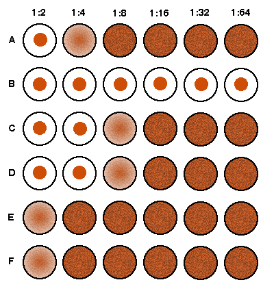
Patient 1 has had more than a four-fold increase in titer, suggesting an acute influenza A viral infection. Patient 2 has a low titer of antibodies that did not increase, so this suggests a past influenza A infection. There is no inhibition in the wells for patient 3, who presumably has never been infected.