

|
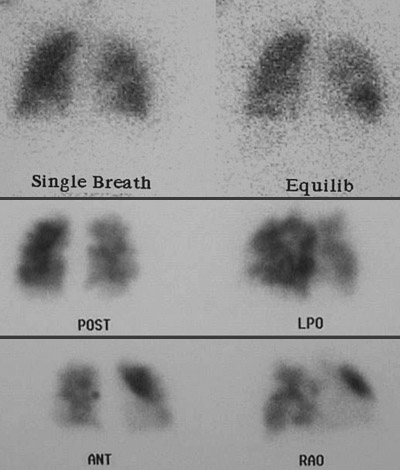
Image 2.1 This is a ventilation-perfusion scan (VQ scan). In the top panel, ventilation is assessed and appears relatively uniform, except for a portion of the left lower lobe. In the bottom two panels, various views indicate multiple areas in which perfusion is diminished, and these areas are different from the area of decreased ventilation. Thus, there is a "V-Q mismatch" that gives a high probability for pulmonary embolism. |
|
|