

|
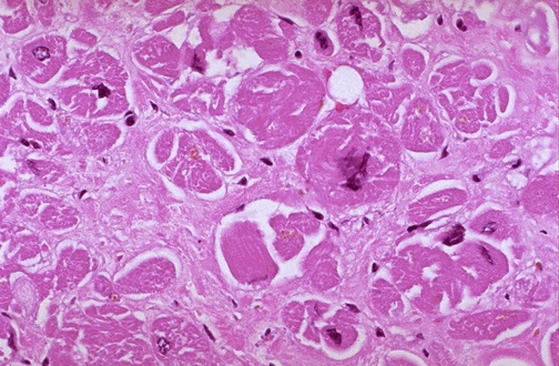
Image 1.2 This is a section of the left ventricular wall. The myocytes are hypertrophied with enlarged, hyperchromatic and irregularly shaped nuclei within the myocytes. Within the interstitium, there is an increased amount of fibrous tissue, but inflammatory cells are not present. In some of these cases one can see rare myofibrillar loss. |
|
|