

|
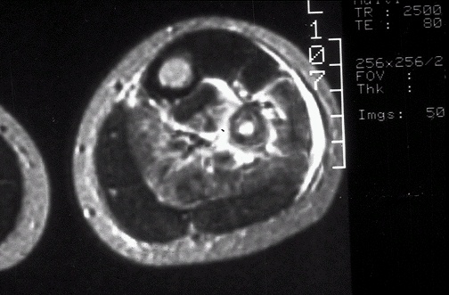
Image 2.2 In this MRI scan in axial (transverse) view through the mid-calf, there is irregular bright extension of the lesion into soft tissue from the bone of the fibula. The normal bone cortex is dark, while normal marrow containing fat is brighter. |
|
|