Image 10
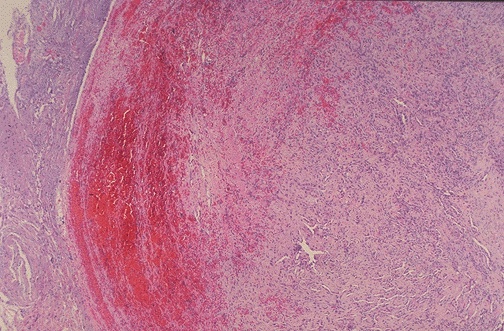
Here is a pelvic vein thrombus seen at low power microscopically. Note the thin muscular wall typical of a vein. The thrombus displays varying degrees of organization, reflecting its propagation over time.