


|
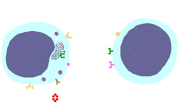
The interaction of a B lymphocyte on the left with a CD4 cell on the right is shown above. The key components of this interaction are labelled below. The microbe is ingested into an endosome and fused with a lysosome to form a phagosome in which the microbe is digested and antigenic peptides produced. The MHC II molecule and the CLIP (class II invariant-chain peptide) molecule are manufactured in the endoplasmic reticulum (ER) and packaged in the Golgi apparatus. The CLIP peptide fills the binding site of the MHC, and when fusion with the phagosome carrying microbial peptide fragements occurs, a microbial peptide displaces the CLIP. The MHC molecule is transported and displayed on the B cell surface. The CD4 lymphocyte then interacts with the B cell, using its T cell receptor (TCR) to recognize the MHC displaying a peptide specific for that TCR. The TCR complex consists of variable alpha and beta chains of TCR that recognize the antigen, along with a gamma chain and a CD3 protein that signal the cell when contact with a specific MHC carrying antigen occurs. Adhesion and signal transduction are further aided by a protein specific to the T cell surface (CD4 in this case). This recognition is aided by a costimulator (B7) on the B cell surface that interacts with CD28 on the T cell surface. This "turns on" the CD4 cell (do they really flash neon pink?) which then uses a CD40 ligand and cytokines to stimulate the B cell and turn on antibody production. This mechanism helps to keep immune responses highly specific for a microbe and produce specific antibody against the microbe. This same interaction can occur between a macrophage and a CD4 cell, leading to macrophage activation for production of costimulators and cytokines that amplify the T cell immune response. |
|
|