- The key to finding problems with the immune system is starting with a proper history and physical examination. It is common for both children and adults to have infections, but they are often minor and recovery is quick. When infections are frequent, prolonged, severe, and are caused by unusual organisms, then an immune disorder is more likely to be present. A physical examination can help determine key organ involvment: is there lymphadenopathy, hepatomegaly, or splenomegaly? Are external lesions present that suggest a particular infection or immune reaction?
- Many of the tests of immune function are serologic tests-a patient serum specimen is utilized. These tests tend to be cost-effective, because they are relatively inexpensive to perform and can yield good information.
- CBC
- The simplest beginning point for assessment of immunologic function is the CBC, which is easy to perform on a blood sample. It is inexpensive. The test is performed on an automated instrument.
- The CBC will provide information about red blood cell numbers and size, leukocyte numbers, and platelet numbers. In addition, many automated instruments can estimate the different types of leukocytes (automated differential count). The traditional method for producing a differential count of the white blood cells is manual-a technician or physician looks at the peripheral blood smear and counts them (100 cell counts are standard).
Coulter Counter
- An increase or decrease in the total leukocyte count will give an indication of what the immune system is doing. In general, an elevated WBC count suggests an acute infectious or inflammatory response. A greatl elevated WBC count suggests a leukemia. A low WBC count suggests a problem with production, often a bone marrow problem.
- Enumeration of the WBCs with a differential count will indicate whether a particular component, such as the neutrophils, is increased or decreased. Remember to multiply the differential percentage by the total WBC count to get the absolute numbers of cells. A WBC differential count with 95% lymphocytes and 5% neutrophils means an absolute lymphocytosis if the total WBC count is 40,000/microliter, but means neutropenia if the total WBC count is 3000/microliter.
- Lymphocyte subsets
- In order to further assess lymphocyte numbers, one can enumerate the CD4 and CD8 cells. This is typically done by flow cytometry. Ordinarily, there are more CD4 cells than CD8 cells in the peripheral blood, giving a CD4:CD8 ratio of greater than 1.
- Quantitative immunoglobulins
- A standard chemistry panel may provide you with an idea of the total amount of serum globulins. If the total protein is 6.6 g/dL and the albumin is 4.5 g/dL, then the difference is globulins. Most of this is gamma globulin.
- However, to be more precise, and to determine the major classes of immunoglobulins present, quantitative immunoglobulin measurement can be performed. Quantitative immunoglobulin measurement includes the three major classes: IgG, IgM, and IgA. An increase of more than one typically implies a polyclonal response to inflammation and/or infection. An increase in one of them may indicate a monoclonal gammopathy which requires further characterization. A decrease in one ore more classes suggests hypogammaglobulinemia with an immunologic disorder involving B cells.
- Electrophoresis
- A serum protein electrophoresis (SPEP) can give a better indication than just a total protein and albumin measurement of the amount of blood proteins present. SPEP is often used as a screening procedure for the detection of various pathophysiologic states, such as inflammation, protein loss, monoclonal gammopathies, and other dysproteinemias.
- Immunoelectrophoresis is used to detect, confirm, and then characterize the presence of a monoclonal gammopathy identified by serum protein electrophoresis (SPEP) in patients suspected of having a plasma cell dyscrasia
- Complement components
- C3 and C4 are routinely measured. C3 and C4 are acute phase proteins that, if elevated, may demonstrate inflammation or infection. Decreased levels may be seen in autoimmune disease, bacteremia, tissue injury, chronic hepatitis, and nephritis. Individuals with congenital deficiencies of early classical pathway components, such as C4, often have lupus-like disorders with arthritis, nephritis, and rashes. Decreased levels may also be seen in systemic lupus erythematosus (SLE), disseminated antigen-antibody complex disease, acute glomerulonephritis, or chronic hepatitis.
- C2 levels can be measured. Increased C2 levels are associated with the acute phase response. C2 deficiency has been associated with increased susceptibility to infection, particularly S. pneumoniae infections, arthritis, rashes, and nephritis. Extremely low levels of C2 may be caused by a genetic deficiency, and half of these cases will have associated autoimmune disease, while half will appear to be normal but have increased susceptibility to bacterial infections.
- C5 is rarely measured. Decreased or absent C5 levels have been associated with systemic Neisseria infections, including recurrent N. meningitidis or N. gonorrhoeae infections, and systemic gonorrhea with arthritis.
- A C1q binding assay is used to detect circulating immune complexes, since C1q can recognize Fc on immunoglobulins.
- C-reactive protein (CRP)
- This is an "acute phase reactant" that binds to polysaccharides present in many bacteria, fungi, and protozoal parasites. Once complexed, CRP becomes an activator of the classical complement pathway. CRP recognizes potentially toxic autogenous substances released from damaged tissues, binds them, and then detoxifies or clears them from the blood. CRP quickly elevates following the onset of infectious and inflammatory conditions. CRP is the most sensitive acute phase protein and will elevate within two hours of acute insult, then peak and begin decreasing within 48 hours if no other inflammatory event occurs.
- Nitroblue tetrazolium test (NBT)
- This is a test of the NADPH oxidase enzyme pathway in peripheral blood neutrophils. In this test, neutrophils on a slide are exposed to a mild stimulus, incubated with NBT, and a 100 cell differential count made. In normal persons, >95% of the neutrophils have a blue-black precipitate in their cytoplasm because of the action of the enzyme, while persons with chronic granulomatous disease have <5% staining.
Nitroblue Tetrazolium (NBT) Slide Test
- Respiratory burst assay
- This test is performed by flow cytometry and can quanitate the degree of NADPH oxidase enzyme activity. Neutrophils are stimulated and the amount of superoxide or hydrogen peroxide generated is measured. Persons with chronic granulomatous disease have reduced amounts.
- Autoantibodies
- There are serologic assays for many different autoantibodies. Some have better sensitivity and specificity than others.
- The antinuclear antibody (ANA) test is the initial screening test used in the evaluation of patients with a suspected collagen vascular disease. A titer is given for positive tests. A titer of 1:160 or greater is considered significant. Titers less than or equal to 1:80 are usually of no or questionable significance, and low titer ANAs are more common with advancing age. The pattern of ANA staining of the substrate (typically a Hep-2 cell line) may have some relationship to disease state. Some generalizations regarding ANA patterns are as follows:
Typical "Homogenous" Pattern of Nuclear Staining of a Positive ANA
- A peripheral pattern of staining suggests anti-DNA antibodies, seen most often in SLE.
"Rim" Pattern
- A homogeneous staining suggests anti-DNA, anti-histone, or anti-deoxyribonucleoprotein (DNP) antibodies observed in SLE, RA, and drug-induced SLE.
- A speckled pattern indicates: 1) Antibody to SSA or SSB, observed in SLE and SS, 2) Smith antibody (Sm) observed almost exclusively in SLE, or 3) RNP antibody observed in SLE, MCTD, RA, and scleroderma.
"Speckled" Pattern
- Anti-nucleolar staining is observed in scleroderma and some forms of Raynaud's phenomenon.
"Nucleolar Pattern"
- A centromere pattern is observed in the CREST syndrome of scleroderma; CREST = calcinosis, Raynaud's phenomenon, esophageal hypomotility, sclerodactly, and telangiectasia
- A variety of additional autoantibody tests can be performed to try and determine the nature of the underlying autoimmune disease. The following table indicates sensitivities of these assays for some of the autoimmune conditions:
|
Autoantibody |
SLE |
Drug-induced SLE |
Sjogren Syndrome |
Rheumatoid Arthritis |
Scleroderma |
PM/DM |
|
ANA |
99% |
99% |
68% |
16-50% |
40-75% |
50-90% |
|
dsDNA |
70% |
1-5% |
1-5% |
1% |
-- |
-- |
|
ssDNA |
80% |
80% |
moderate |
moderate |
-- |
-- |
|
Smith |
30% |
1% |
1-5% |
1% |
<1% |
<1% |
|
SSA |
25-35% |
low |
10-70% |
low |
-- |
low |
|
SSB |
15% |
low |
15-60% |
low |
low |
-- |
|
RNP |
50% |
-- |
5-50% |
5% |
20% |
-- |
|
Scl-70 |
-- |
-- |
5% |
-- |
20-60% |
5% |
|
Jo-1 |
-- |
-- |
-- |
-- |
-- |
30-50% |
- As can be seen from this table, there is plenty of overlap among the various autoimmune conditions, and not all cases will be detected. Thus, clinical correlation is necessary for interpretation. These serologic tests are helpful, but not diagnostic.
- Serologic assays for infectious diseases
- The diagnosis of many infectious diseases can be made or confirmed by the presence of antibodies to a particularly microbial agent. Most infections will elicit some sort of antibody response, though this response may not be the body's main defense against the organism.
- Serologic assays for infectious agents are often divided into separate assays for IgM and IgG antibody. In general, the presence of an elevated IgM titer suggests a recent infection. The presence of an elevated IgG titer suggests that infection at some point has occurred. Rather than a single measurement, "acute and convalescent" titers can be done. The increase in an overall titer (mostly IgG) of four fold in two weeks' time indicates recent infection.
- Bone marrow biopsy and aspirate
- This procedure typically samples marrow from the posterior iliac crest in adults. The sternum can be sampled, but only for an aspirate. This procedure is typically done when a malignant process involving the marrow is suspected (leukemia, lymphoma, metastases). In some cases of suspected systemic infection, the marrow may be sampled for diagnosis or for determining treatment. In cases of suspected immunologic disorders, the marrow may be sampled to determine the nature of the cell populations present and to obtain cells for further analysis (typically genetic analysis).
- Lymph node biopsy
- In cases of lymphadenopathy in which a malignant process is suspected, a lymph node may be obtained. Less frequently, nodes are obtained in cases of suspected infectious processes. In general, nodes are not sampled in cases of immunologic disease, because the lymphoid population can be evaluated on the basis of lymphocytes in the peripheral blood circulation.
- Skin biopsy
- Light microscopy and immunofluorescence studies are occasionally done for autoimmune conditions that may involve the skin.
- Laboratory Testing Principles
- Results fall into four categories:
- True positives (TP) Persons who really have the disease and test positive
- False negatives (FN) Persons who really have the disease but test negative
- True negatives (TN) Persons who do not have the disease and test negative
False positives (FP) Persons who do not have the disease but test positive
- The usefulness of a laboratory test can be measured by:
- Diagnostic Sensitivity: how well can the test detect persons who really have the disease?
-
Sensitivity = true positives ÷ (true positives + false negatives)
- Diagnostic Specificity: how well can the test exclude persons without the disease?
-
Specificity = true negatives ÷ (true negatives + false positives)
- Example: In a given population, 1000 persons are tested for the presence of a particular disease. Of these, 80 are found to test positive. However, only 40 of these are found on subsequent confirmatory testing to really have the disease. Furthermore, follow-up of the original group of patients reveals that there were 10 people who really had the disease, but were missed by the initial screening test. Calculate the diagnostic sensitivity and specificity for the original screening test:
-
Sensitivity = 40 true positives ÷ (40 true positives + 10 false negatives)
-
= 80%
-
Specificity = 910 true negatives ÷ (910 true negatives + 40 false positives)
-
= 96%
- So what does a positive or negative test really mean? This can be measured by positive and negative predictive values (PV):
-
PV of a positive test = true positives ÷ (true positives + false positives)
-
PV of a negative test = true negatives ÷ (true negatives + false negatives)
- In the example of the screening test above:
-
PV(+) = 40 true positives ÷ (40 true positives + 40 false positives)]
-
= 50%
-
PV(-) = 910 true negatives ÷ (910 true negatives + 10 false negatives)
-
= 99%
- Predictive values have a lot to do with the prevalence of the disease, or the number of persons in the population who actually have the disease (incidence of disease is only the new cases that are reported). In the above example, the prevalence of the disease was 6%, which is quite high. Few diseases have that high a prevalence in a population.
The prevalence of most diseases is low. Thus positive predictive value, even for a good test with a sensitivity of 95%, can be poor when there are few persons with the disease, and most of the positives will be false positives.
- As an example, the best test in the laboratory is the HIV antibody test, which has a sensitivity of 99.9% and a specificity of 99.7%. In a given population (such as in rural areas) where the prevalence of the disease being tested is around 1:10,000 the predictive value of a positive test will be quite low. Of course, the test is still useful, but it is a screening test, and a repeat assay and an additional confirmatory test (Western blot) are needed to find the true positives.
- The following chart indicates the performance of testing based upon prevalence:
| Prevalence of Disease (%) |
Predictive Value of a Positive Test (%) |
| 1 |
16 |
| 2 |
28 |
| 5 |
50 |
| 10 |
68 |
| 25 |
86 |
| 50 |
95 |
- The generalist, primary care physician is the initial person who sees many patients and who has to deal with the problem of ordering and interpreting screening tests.
- Why can't you have both 100% sensitivity and 100% specificity? The ranges of test values in a population typically have some overlap for persons with and without the disease:
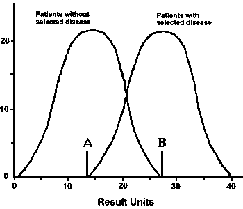
- You can obtain maximum sensitivity at point A, but only at the expense of generating many more false positives that require additional workup to exclude.
- You can obtain maximum specificity at point B, but only at the expense of generating many more false negatives and miss patients with the disease.
- You can improve predictive value by first narrowing down the population to be tested with standard history and physical exam (e.g., don't order superfluous lab tests). Example: you can progressively improve you chances of getting a meaningful result for a prostate specific antigen test if you order it on: men (this should be obvious), older men, older men with a palpable nodule.
- Performance Characteristics of Laboratory Testing
- Accuracy: How well does the test measure what is really there? Agreement of the test results with the patient's condition is the best measure of accuracy.
- Example: clinical diagnosis of acute appendicitis is about 90-95% accurate
- Question: How accurate is the standard history and physical exam?
Question: What is the "gold standard" by which you measure accuracy? Is it the word of your attending physician? A consultant? A laboratory test result? Autopsy? (Note: a courtroom decision on a medical matter may not be based upon scientific principles, but nonetheless can modify how we practice.)
Example: you perform a physical examination on a newborn and determine that the baby has slanting epicanthal folds, bilateral transverse palmar creases, and an absent distal flexion crease on the fifth digits of both hands. You suspect Down syndrome. The "gold standard" is cytogenetic analysis of baby's cells, which reveals a 47, XY, +21 karyotype.
- Precision: How reproducible is the test under the same conditions? The laboratory tries to assure reproducibility by the use of control specimens with each run of patient specimens. The instruments have a routine maintenance and check procedure performed as well.
- You can be precise but not accurate by making the same error consistently.
Example: you may be using improper technique to measure blood pressure, but you will keep getting the same result, which is different from what the nurse (who is positioning the cuff properly and listening appropriately) records.
- Accuracy and precision can apply to written and verbal communcations. Lack of understanding or failure to properly record observations can have an impact.
- Example: the patient's primary physician palpates a "lump" on the left side of the neck of his 17 year old patient. An imaging study is performed, and the lesion is a 3 cm well-circumscribed cyst in the soft tissue of the left lateral neck. The lesion is recorded as consistent with a "brachial cleft cyst". The surgeon's operative report records removal of a "brachial cleft cyst". The surgical specimen is sent to pathology, and the final reported diagnosis is "brachial cleft cyst". The resulting medical record is quite precise, but totally innaccurate, because everyone has made the classic freshman anatomy mistake of confusing the terms "brachial", "branchial", and "bronchial". (It is a branchial cleft cyst.)
- CV: Coefficient of variation. Just how variable are the test results. This depends upon the test methodology, the instrument being used, and the range of results. (The CV is calculated by dividing the standard deviation by the mean.)
- Examples:
- Sodium (Na) of 138 mmol/L is probably between 137.5 and 138.5 mmol/L
- Hgb of 10.0 g/dL is probably between 9.8 and 10.2 g/dL
- Glucose of 800 mg/dL is probably between 770 and 830 mg/dL
- Thus, a change in values from one day to the next generally has to be 10% or more to be of major significance. Just specimen handling, processing, and instrument variation can account for some changes. Running a test in duplicate will show this.
- Bear in mind that there is also "physiologic variation" in patients that is dictated by factors such as the degree of hydration, diet, and exercise.
Example: an elderly person admitted with an apparently normal hemoglobin, but an elevated urea nitrogen and glucose, may be dehydrated, and upon administering fluids will be found to have anemia, but normal renal function, and the glucose was slightly high because she just ate.
- If you rely on specific "numbers" for decision points, you may run into trouble.
Example: it is late afternoon and the physician checks lab values for tests ordered on his patients. He notes that his elderly patient has a hemoglobin of 9.9 g/dL, whereas the value was 10.1 g/dL early in the morning that same day. The physician's "set point" for ordering a transfusion is 10 g/dL, even though this is not a recognized practice standard. Using such criteria, an unnecessary transfusion, subjecting the patient to potential complications, would be given. But the two values could have come from either the morning or afternoon specimens run in duplicate!
- What is "normal"?
- The laboratory sets "normal" ranges for laboratory tests based upon population studies. A test may have a single normal range, or there may be different normal ranges based upon age, sex, race, or other factors. Sometimes, more history is needed for interpretation (such as with maternal serum alpha-fetoprotein in pregnancy, which is dependent upon the gestational age--the later in gestation, the more AFP is present normally) so that is why this information needs to be provided. Otherwise, you may have an uninterpretable result.
- Standard "normal" ranges for tests with numeric values are based upon use of a bell shaped curve. "Normal" is defined as those test values that fall within 2 standard deviations of the mean, which includes 95% of all results. The standard deviation is just a measure of dispersion.
Thus, there is a 1 in 20 chance that an "abnormal" test may really be normal. If you perform 20 or more independent tests (which is not uncommon on patients admitted to hospital), then there is a greater than 50% likelihood that one or more tests will be "abnormal" just from statistical variation. If you keep ordering more tests just to track these down, you can go on for a long time and spend a lot of money.
However, size counts! The farther out of range the test result is, the more likely that the result reflects real disease.
- GUIDING RULE: It is better to treat the patient than the numbers.
- What are the accuracy, precision, and preditive values for clinical assessment?
- Very little may sometimes be done in regard to quality assessment of clinical activities, such as history taking and physical examination. The following story illustrates this point:
- In 1888, Nellie Bly (Elizabeth Cochrane) was a reporter for the New York World, the premier tabloid of its era. She was one of the first true investigative reporters, although a lot of what she did was publicity stunts to sell newspapers (such as her most famous stunt, "Around the world in 80 days" which was made in 72 days, 6 hours, and 11 minutes). One of her stunts that served a useful purpose was an exposé of the New York mental health care system, which consisted of asylums where the mentally ill were placed. She acted the part of an insane woman and allowed herself to be committed to Blackwell's Island, New York City's most notorious insane asylum. She then wrote an exposé of the mistreatment of patients that got the attention of reformers and readers alike, shown in the front pages of the New York World, and that got the asylum closed down. She described the asylum as "…a human rat-trap. It is easy to get into the place, but once you are there, it is impossible to get out." In fact, the editor of the newspaper had to get the police to extricate her 10 days later from the asylum. The diagnostic tools and criteria employed were so poor that the staff could not, or would not, determine who was really mentally ill and who wasn't.
- Medical Necessity
- When you order tests or procedures, you must document the medical necessity for the order (i.e., you must justify what you are doing). Failure to do so will result in the charges for the test or procedure being denied (i.e., you or the institution for which you work will not get paid).
- If you order tests based upon misinterpretation of findings from previous testing, the problem is compounded.
- Every test ordered must have a reason. Charges for tests which have documentation that indicates they were done as "standing orders" or as "routine" will be flatly denied.
- Tests may be appropriate depending upon the time course of a workup for disease. Primary physicians may appropriately order screening tests. However, if a urologist were to order a "screening prostate specific antigen test" then the charge would be denied.
- COCHRANE'S APHORISM: Before ordering a test, decide what you will do if the test is 1) positive, or 2) negative. If both answers are the same, do not order the test.
- Role of Epidemiology
- Case Presentation:
- History: A 32 week gestational age fetus was stillborn to a 23 year old mother who had been experiencing a low grade fever with mild diarrhea for a couple of weeks. She did not feel fetal movements for about a day and went to see her doctor, who could not hear fetal heart tones, and no fetal cardiac activity was noted on ultrasound. The fetus was stillborn and was observed to be slightly hydropic.
Autopsy: The fetus was found to be consistent with 32 weeks gestational age. Hepatosplenomegaly was present. Spleen, liver, and lungs showed small yellowish-tan, pinpoint granulomas (or microabscesses). These lesions were also present in the placenta. The brain showed a meningitis. A blood culture was taken at autopsy. The culture grew Listeria monocytogenes.
Epidemiology: The above findings were reported to the health department. The health department tracks such cases over time. They noted that there was an increase in other reported cases of this illness during that same month (May).

- What are the next steps that the health department should take?
- A slight seasonal variation in cases is noted, but there is a marked increase in cases in May of the current year that suggests more than just sporadic cases. Affected persons were interviewed to determine the circumstances of the infection.
- Serologic studies were done on the isolates of Listeria from culture to determine if they were all of the same serotype, which would suggest a single source.
- What do you think might be the source of this increase in cases?
- Listeria has the ability to survive for long periods of time in soil, water and on vegetation. Thus, it is likely to be a food or water-borne contaminant. L. monocytogenes is a short, gram positive, pleomorphic organism that is non-spore forming. It is a non-capsulated motile rod that grows in a temperature range of 4 to 44°C, with an optimum temperature of between 25 and 36°C. Thus, L. monocytogenes is prevalent in cooler temperatures between the months of November and April (northern hemisphere) and seldom occurs during hot months or in areas of the world that have year-round hot climates. This can be explained by the psychrophilic characteristics of the organism.
- Eleven L. monocytogenes serotypes have now been described. Of these, Types 4b, lb and la account for most of the human cases in the US. Neonates comprise the largest group with Listeriosis infections, followed by an increasing number of cases with advancing age groups. Listeriosis may be found in apparently healthy, asymptomatic persons.
- Listeriosis is generally a low-grade infection in adults, but can be more severe in pediatric populations, and it can be a congenital infection that can lead to fetal demise. Interview of the affected adults revealed that they had all consumed a certain brand of cheese. The manufacturer of the cheese product was found to be using unpasteurized milk, and the factory was closed after it was determined that the Listeria cultured from the milk storage tanks was the same serotype as for the affected adults (4b).
|
 Return to the Immunology Course Outline