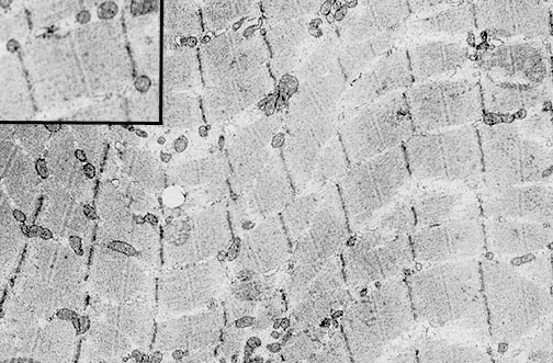
| In this electron micrograph of normal skeletal muscle, myofibrils are present, surrounded by an endomysium with vascular supply and peripheral nuclei. Each myofibril is composed of interdigitating thin actin and thick myosin filaments. The inset at the upper left demonstrates a single sarcomere with thin actin and thick myosin filaments that interdigitate, bounded on each side by a dark Z disc, and a central M line. |