


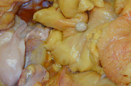
| The small, round object seen here in the lower pelvis next to the uterus at the lower left is a so-called "peritoneal loose body" which was derived from an appendix epiploica (a tag of adipose tissue on the surface of the colon). They may be attached by a thin pedicle that undergoes torsion, leading to infarction, typically in older persons. This may cause a condition known as primary epiploic appendagitis (PEA), a condition that can mimic diverticulitis or appendicitis. Clinical features of PEA, an uncommon condition, include sudden onset of focal abdominal pain in an otherwise healthy patient with mild or absent secondary signs of abdominal pathology. Computed tomographic findings of PEA may include: a 1 to 4 cm ovoid paracolic lesion with fat density and adjacent fat stranding, bowel wall thickening and/or compression, and thickened visceral and/or parietal peritoneum. There is often a central high-attenuating dot because the infarcted tissue tends to calcify. This small calcified body may become detached, as in this case, and appear as a "peritoneal loose body" or "peritoneal mouse" in the abdominal cavity. It may re-attach itself to a surface, such as the lower aspect of the spleen, in which case it can be called a "parasitized appendix epiploica." The smooth surface and hard, calcified consistency of the appendix epiploica help to distinguish it from a metastasis. |
|
|