


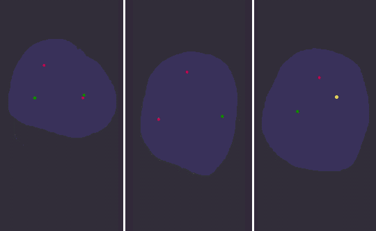
| The FISH technique is shown above with interphase nuclei in three panels. A specific DNA probe with a fluorescent tag identifies a specific region of a chromosome. Different colored fluorescent tags for probes allow identification of various abnormalities. In the left panel, probes specific for the breakpoint regions of chromosome 8(q22) in green and chromosome 21(q22) in red identify a translocation t(8;21) in a patient with acute myelogenous leukemia. In the center panel, probes to chromosomes 11 (green) and 13 (red) identify a deletion of 11q23 in a patient with small lymphocytic lymphoma. In the right panel, a red fluorescent tag to the BCR gene of chromosome 22 and a green tag to the ABL gene of chromosome 9 are seen, but the yellow dot identifies the abnormal BCR-ABL fusion gene in a case of chronic myelogenous leukemia with the "Philadelphia chromosome". |
|
|