Purpose: To evaluate the feasibility and efficiency of MRI in assessing the delivery, colonization and survival of stem cells after direct injection into infracted and normal myocardium of rat.
Methods: Rat mesenchymal stem cells (MSC) were labeled with Feridex (ferumoxides). MI was induced in rats by ligation of the left anterior descending coronary artery. Labeled MSC were injected to MI and sham-MI rats. MRI was done after 7, 14 and 30 days from cell transplantation. In another group, as a control, BrdU labeled MSC were injected to MI rats. At the end of the study the rats were sacrificed and histochemical studies were done for the presence of MSC in the heart and other tissues.
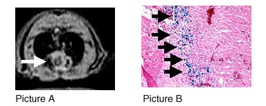
Results: Ferumoxides wass incorporated into MSC in culture. Phantom MRI of labeled cell suspension demonstrated a significant decrease in signal intensity due to incorporated iron, in relation to unlabeled cell suspension. In vivo T2 weighted, gradient-echo MRI of injected rats demonstrated well-defined left ventricular signal voids in the area of cell injection (Picture A, arrow). Those susceptibility artifacts persisted for at least 24 days. No artifacts could be seen in rats injected with unlabeled (control) cells. Prussian-Blue staining of tissue sections has proved the presence of iron-laden blue cells in injected animal hearts (Picture B). In another arm of the study, the use of poly-L-lysine as a transfection agent in a mixture with ferumoxides, greatly increased the uptake of the iron particles by MSC.
Conclusion: We have shown, for the first time that monitoring the fate of transplanted stem cell by MRI is feasible in rat. Our work provides a new technology for in vivo, long-term, non-invasive tracking of implanted cells by a commercially available contrast agent.