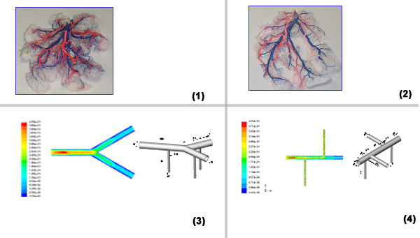
Fetal development is largely dependent on adequate feto-placental perfusion, which is a function of the vasculature anatomy. The gross anatomy of the feto-placental vasculature reveals three groups: chorionic vessels, intraplacental (IP) vessels and capillaries where gazes and nutrition exchange take place. The chorionic vessels are superficial and distributed across the chorion in two major patterns, dichotomous and monopoidal. Ten placentas were delivered at 38-41 weeks of gestation where the babies weights from 2.700 Kg to 3900 Kg. The cast model allowed observing the whole vasculature anatomy of the placenta (Figs. 1 &2): insertion of umbilical vessels, chorionic and IP vessels and the capillary system. The chorionic vessels had about 6-8 generations (bifurcations) from the insertion towards the margins of the chorionic plate where the diameter of the last generation ranges from 0.16 to 1.89. The chorionic vessels were distributed in a mixed pattern of dichotomous and monopoidal. In the monopoidal pattern the diameter of the main vessel remained fairly constant (10% change in the diameter). The IP vessels were surrounded by thick system of capillary vessels, as a consequence, only those who were fairly bare were measured. The IP vessels entered the placenta at angle 60-90° where their diameters ranges from 0.33 mm to 0.93 mm. The IP vessels were always thinner than the chorionic vessels, with a ratio 0.16-0.77.
The computational simulation of the feto-placental blood circulation was done by solving the Navier Stokes equations in two simplified models that represent typical segments of dichotomous and monopoidal networks of the chorionic arteries (Figs. 3&4). The velocity at the inlet of both models was 0.32 m/s. The velocity field in the chorionic vessels decreased when the vessels split. However, the velocity in the IP vessels was 1.75 m/s, regardless the velocity field in the mother vessels. In summary, our simulations indicated that the pattern of the chorionic vessels did not affect the velocity magnitudes that were developed within the IP vessels in the dichotomous or monopoidal models. In order to clarify this point, it is necessary to make additional experiments and simulations of complex geometry of the placenta.