
Objective: to follow the changes of VEGF and iNOS expression in the intact and infarcted rat heart post LELI. Estimation of newly formed capillaries Methods: Two animal models were used: (1) The left anterior descending (LAD) coronary artery was ligated in rats to create MI. The hearts of laser irradiated (LI) rats received irradiation following LAD occlusion and were sacrified at various time intervals (control rats did not recive irradiation). (2) Intact hearts were irradiated (the radiation was applied through the intercostal muscles) and rats were sacrified after 7 days. Homogenates from the hearts were analysed for VEGF and iNOS by western blot analysis. A densitometer software (Image master1D prime ver.3.01) was used to determine optical density of the blots.Brdu kit was used for the estimation of newly formed capilaries.
Results: VEGF content was measured in infarcted hearts that were sacrified 2.5 hours, 1, 2 and 3 days after the occlusion. In the LI hearts at 2.5h and 1 day time intervals there was not increase in VEGF content as compered to non irradiated ones. In 2 and 3 days post MI the content of VEGF was significantly elevated (2.4-fold and 1.35-fold respectivly) in the LI rats as compered to non-irradiated. The content of iNOS was significantly increased (2.7-fold) in LI hearts 3 days post MI as compared to non-irradiated hearts. In the intact irradiated hearts VEGF content was significantly higher (2.8-fold) then in the non-irradiated ones.
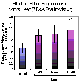
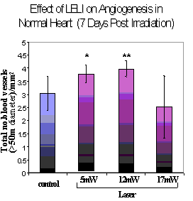
After irradiation of intact hearts with 5mW, 12mW and 17mW a significant increase (of 1.56, 1.6 and 1.82 folds, respectively) was observed in the amount of newly formed capliaries when compared tountreated hearts.
Conclusion: It is concluded that LELI markedly increased the content of VEGF and iNOS in the infarced heart. An elevation of VEGF and iNOS in infarcted heart is significant for angiogenesis and the maintenance of the blood flow. The elevation of VEGF by LELI in the non-infarcted hearts may indicate the possibility of protecting and enhance angiogenesis. According to the Brdu results there is formation of new blood vessels after irradiation.
 |
 |
